
Figure 4. Validation of ZSCAN1 autoantibodies in CSF and sera of ROHHAD patients using 293T Cell-Based Assays.
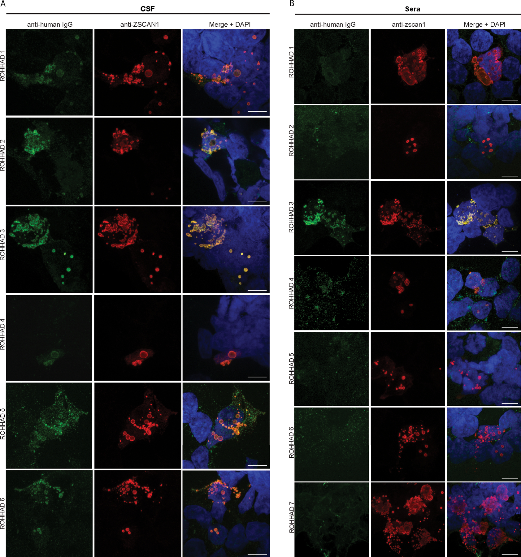
(A) CSF: Immunocytochemistry with 293T cells expressing full-length ZSCAN1 and immunostaining with CSF (1:10) from ROHHAD patients and commercial antibody to ZSCAN1 (Rabbit, 1:1000 Invitrogen). Anti-human IgG-488 was used to visualize human IgG and anti-Rabbit IgG-567 was used to visualize anti-ZSCAN1 commercial antibody. Exposure times and post-image processing and thresholding was kept constant across conditions within the experiment. Co-localization was assessed qualitatively through observance of yellow in merged images. (B) Sera: Immunocytochemistry with 293T cells expressing full-length ZSCAN1 and immunostaining with Sera (1:100) from ROHHAD patients and commercial antibody to ZSCAN1 (Rabbit, 1:1000 Invitrogen). Anti-human IgG-488 was used to visualize human IgG and anti-Rabbit IgG-567 was used to visualize anti-ZSCAN1 commercial antibody. Exposure times and post-image processing and thresholding was kept constant across conditions within the experiment. Co-localization was assessed qualitatively through observance of yellow in merged images. Note colocalization in ROHHAD Sera-1 and 3.