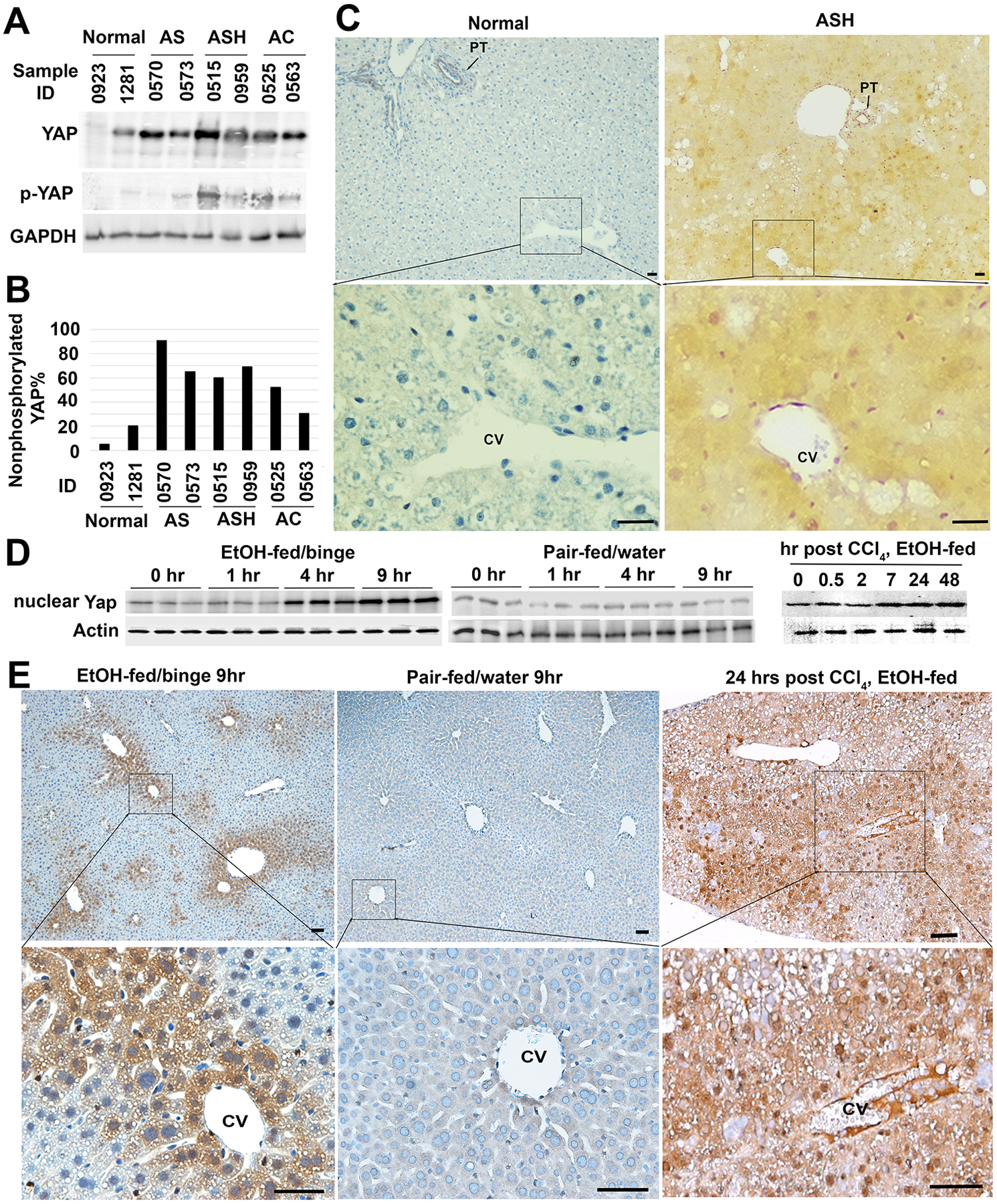
Figure 1. YAP activation during progression of alcohol-related liver disease and in experimental models of alcohol-associated liver injury.

(A) Total YAP and its phosphorylated form at serine 127 residue in six patients with alcoholic liver disease were examined with specific rabbit antibodies in Western analysis. (B) Densitometrical analysis was performed to calculate percentage of the active nonphosphorylated YAP based on band intensity of phosphorylated (inactive) and total forms of this protein in (A). AS: alcoholic steatosis; ASH: alcoholic steatohepatitis; and AC: alcoholic cirrhosis. (C) IHC detected YAP accumulation in ASH livers. Scale bar: 50 μM. (D) Immunoblotting and densitometry analyses showed Yap accumulation in nuclear fraction of 5% ethanol-fed animals that received ethanol binge (EtOH-fed/binge), but not in pair-fed livers that received water gavage (pair-fed/water). Accumulation of nuclear Yap was also found in livers that were exposed to moderate ethanol and one acute dose of CCl4 intoxication. Quantification was performed based on three independent experiments from 5 animals per group. *P<0.05. (E) IHC detected abundant Yap protein in pericentral hepatocytes of ethanol-fed livers at 9 hours after binge or 24 hours post CCl4 intoxication. Scale bar: 100 μM.
