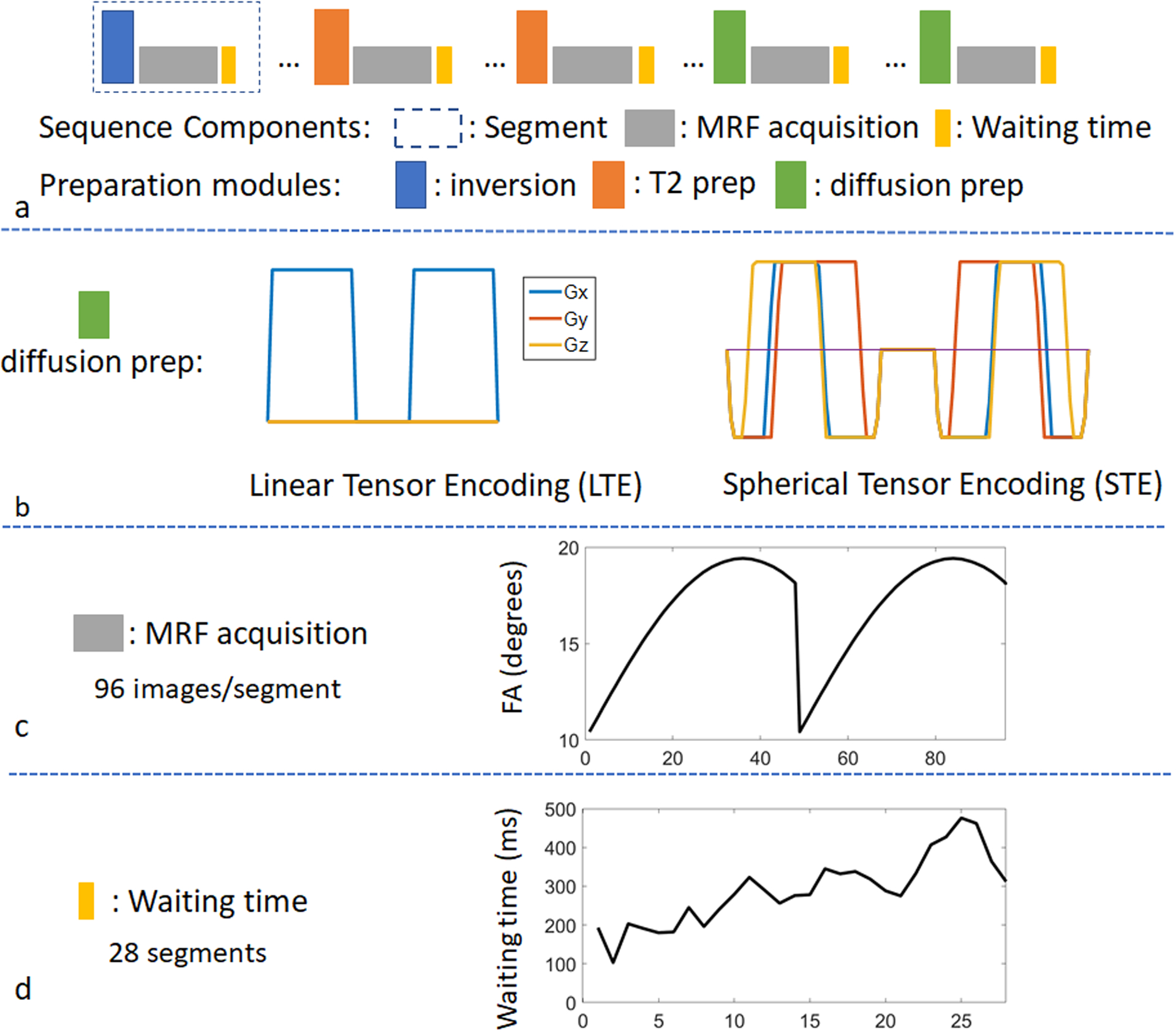
Figure 1.

The sequence structure of a multidimensional MRF (mdMRF) scan. (a) acquisition segments, including T1 inversion pulses, T2 preparations, diffusion preparations and MRF readouts. (b) the gradient waveforms for linear and spherical tensor encoding. (c) an example of a flip angle pattern used in the mdMRF scans and (d) an example of the waiting time pattern added at the end of each segment.
