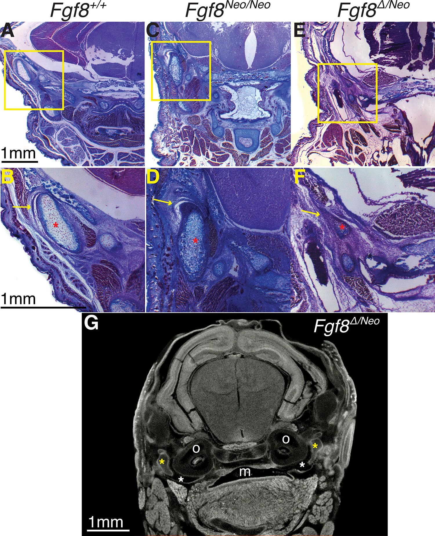
Figure 6: Severe reductions of Fgf8 deform the temporomandibular jaw joint (TMJ).

Trichrome stained coronal sections of Fgf8+/+ (WT; left column), Fgf8Neo/Neo (middle column), and Fgf8Δ/Neo (right column) neonatal (P0) skulls. Yellow boxes in upper panels are shown magnified in lower panels. A,B) In WT, the temporomandibular joint (TMJ) has a joint disc (yellow arrow in B) below a defined glenoid fossa creating an upper joint cavity. C,D) The condyle in Fgf8Neo/Neo mice is dysmorphic and rotated vertically and the upper joint cavity is reduced, but an articular disk (yellow arrow in D) is still present. E,F) Fgf8Δ/Neo neonates have a severely reduced glenoid fossa and condylar process, and the joint cavity and articular disc are absent (yellow arrow in F). Red asterisks highlight the condylar process. G) micro-CT scans of Fgf8Δ/Neo show the oral cavity opening into the tympanic cavity (white asterisk). Yellow asterisks highlight the malleus. mouth (m), otic capsule (o).
