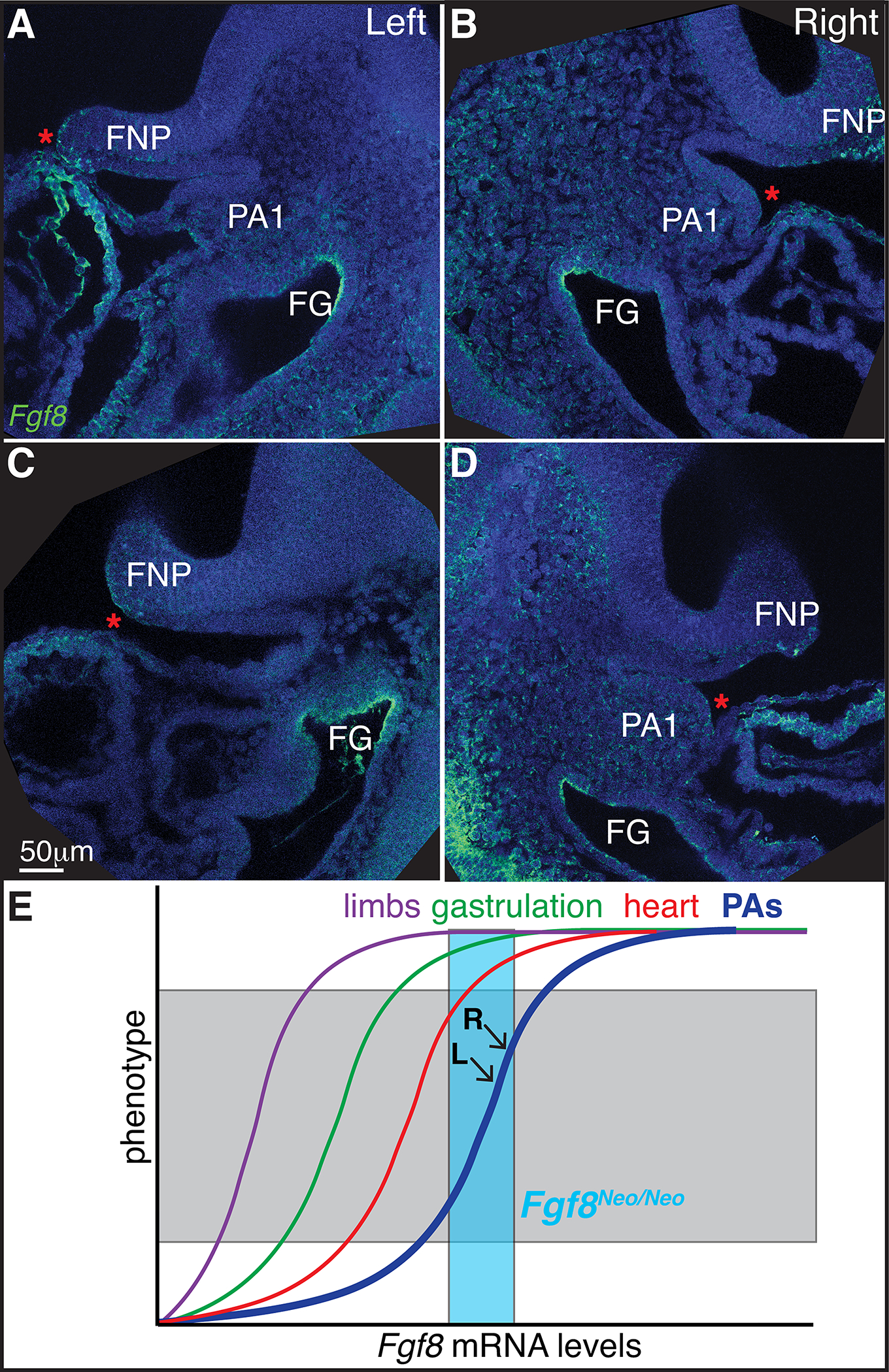
Figure 8: Fgf8 is expressed asymmetrically in the developing heart.

A-D) Saggital sections of fluorescent in situ hybridization of Fgf8 (green) in E9.5 embryos. Left (A,C) and right (B,D) sides are shown for two separate specimens. Sections shown are those where the developing heart tube comes closest to the first pharyngeal arch (PA1) for each side. Nuclei are counterstained with dapi (blue). E) Model for non-linear relationships for Fgf8 levels and tissue morphogenesis. Teal box represents Fgf8 levels in Fgf8Neo/Neo mice. L and R indicate levels in left and right sides of PA1 (see text for more details). Frontonasal process (FNP), foregut (FG).
