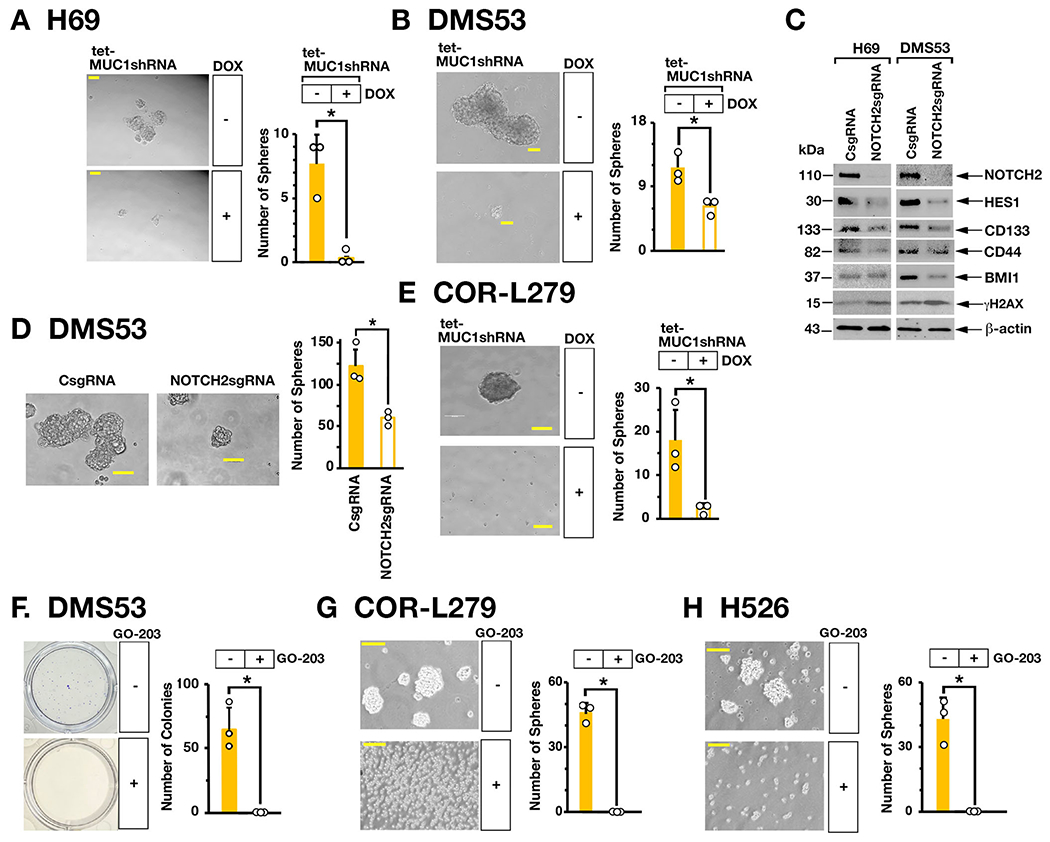
Figure 5. MUC1-C is necessary for SCLC self-renewal.

A and B. Representative images of tumorspheres derived from H69/tet-MUC1shRNA (A) and DMS53/tet-MUC1shRNA (B) cells treated with control vehicle or DOX for 7 days (left). Bar represents 50 microns. The number of tumorspheres is expressed as the mean±SD of three determinations (right). C. Lysates from H69 (left) and DMS53 (right) cells expressing a CsgRNA or NOTCH2sgRNA were immunoblotted with antibodies against the indicated proteins. D. Representative images of tumorspheres derived from DMS53/CshRNA and DMS53/NOTCH2shRNA cells (left). Bar represents 50 microns. The number of tumorspheres is expressed as the mean±SD of three determinations (right). E. Representative images of tumorspheres derived from COR-L279/tet-MUC1shRNA cells treated with control vehicle or DOX for 7 days (left). Bar represents 50 microns. The number of tumorspheres is expressed as the mean±SD of three determinations (right). F. DMS53 cells treated with control vehicle or 5 μM GO-203 for 3 days were seeded in dishes for 12 days. Representative images are shown for colonies stained with crystal violet (left). The number of colonies is expressed as the mean±SD of three determinations (right). G and H. Representative images of tumorspheres derived from COR-L279 (G) and H526 (H) cells treated with control vehicle or 5 μM GO-203 for 7 days (left). Bar represents 50 microns. The number of tumorspheres is expressed as the mean±SD of three determinations (right).
