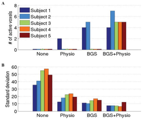
Figure 2.

A: Number of activated voxels detected in each subject (sum of the left and right LGN voxels) and B: Standard deviation of the estimated residual noise in LGN voxels when no correction methods were applied (first group on left), only physiological noise reduction was applied (2nd group), only background suppression (BGS) was applied (3rd group), and both physiological noise reduction and BGS were applied (4th group). The largest improvement in the sensitivity of the detection occurred when both BGS and physiological noise reduction were used. A value lower than 1 in (A) indicates no active voxels were found. [Color figure can be viewed in the online issue, which is available at www.interscience.wiley.com.]
