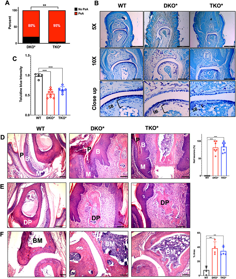
Figure 2: Psoriatic-arthritic-like (PsA) phenotype in mice with severe psoriasis-like disease.
A. Prevalence of psoriatic arthritis (PsA) in mice with inducible dual epidermal deletion of c-Jun and JunB (DKO*) and triple epidermal deletion of c-Jun, JunB and S100A9 (TKO*) with severe psoriasis-like disease (n=20 per group). B. Toluidine blue staining of the distal interphalangeal (DIP) joint of control wild-type (WT), DKO* and TKO*mice with PsA (scale bar =200μm). C. Quantification of toluidine blue staining intensity of articular cartilage in WT, DKO* and TKO* mice (WT n=4; DKO* n=8; TKO* n=6; each point represents the median of several joints measured per sample). D. Hematoxylin & Eosin (H&E) stained histological images showing psoriatic nail involvement with changes in the nail plate (P), nail matrix (M) and nail bed (B) of DKO* and TKO* mice and quantification of nail lesions (Scale bar=100 μm). E. H&E histological images of the distal phalanx (DP) showing enthesitis with high immune infiltration in the areas around the bone (Scale bar=100 μm). F. H&E histological images showing osteitis of the bone marrow (BM) of the distal phalanx (DP) in DKO* and TKO* mice (BM=bone marrow) with quantification of percent area of bone marrow covered by inflammation (Scale bar=100 μm).

