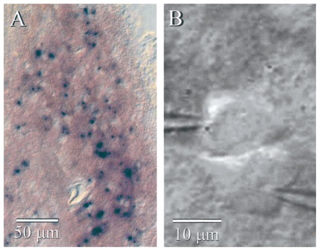
Figure 1.
IO neurons from KO mice. A, β-Galactosidase histochemistry of a parasagittal section from a Cx36 KO mouse shows many strongly stained cells in the IO nucleus. B, IR-DIC image of two very closely spaced neurons during paired whole-cell recordings from the IO of a KO mouse. Despite their proximity, these cells were not electrically coupled to each other.

