
Figure 2. Diffusion ranges of injury-released VEGF and FGF.
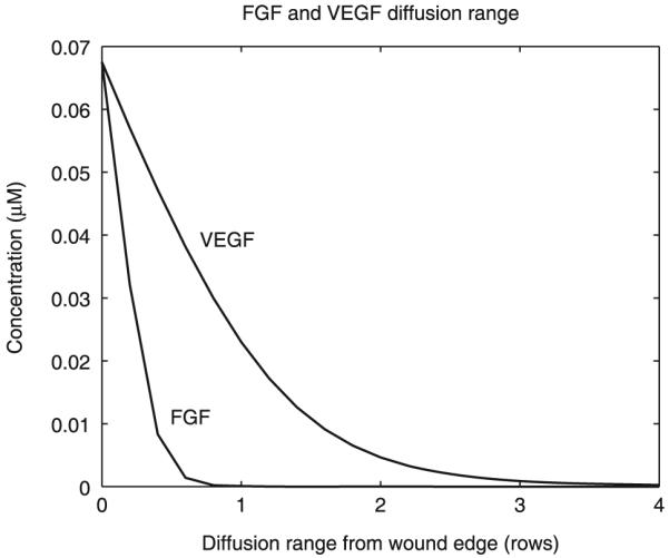
Diffusion Ranges are predicted by the mathematical model and are plotted as number of cell rows from the wound edge at 60 s after injury. FGF, fibroblast growth factor; VEGF, vascular endothelial growth factors.