
Figure 7. Wound closure rate of HUVECs transfected with PLCδ-containing plasmid and empty plasmid.
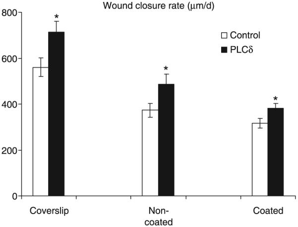
Cells were seeded on glass coverslip, non-coated, and gelatin-coated tissue culture dishes. Wound closure rate is significantly higher in PLCδ-transfected cells grown on all three different surfaces (*P < 0.01, n = 8). HUVEC, human umbilical vein endothelial cells; PLC, phospholipase C.