
Figure 1:
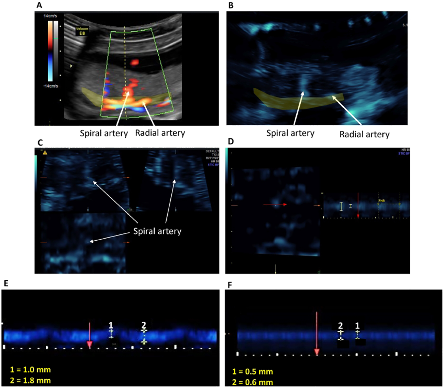
Illustration of the B-Flow/STIC M-mode technique. A spiral artery was identified in the placental bed (yellow shaded area) using power Doppler (Panel A). B-flow was activated and the spiral artery was demonstrated at the same anatomical location (Panel B). A 4D- STIC block was acquired and the spiral artery was visible in 3 orthogonal planes (Panel C). M-mode was activated at the Z-axis and vessel luminal diameter was measured perpendicular (red arrow) to the vessel lumen (Panel D). Fetal heart rate (FHR) was measured each time to confirm that the blood vessel was a maternal vessel. Panels E and F show representative B-flow/STIC M-mode measurement of luminal diameters of two different spiral arteries during the cardiac cycle (1: diastole, 2: systole) in an untreated (E) and SAR suppressed (F) baboon.