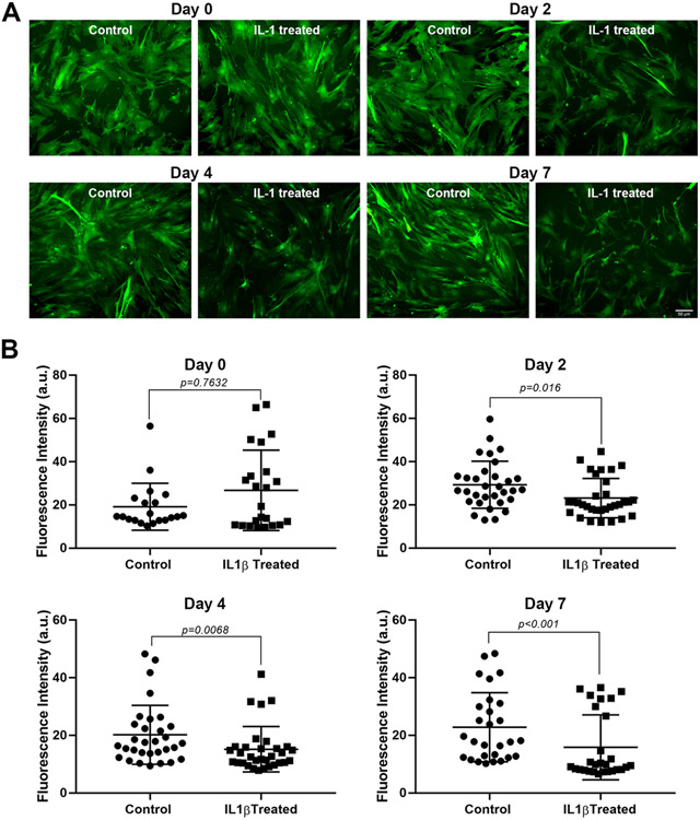
Figure 1. Effect of IL-1β on GFP intensity.
Lacrimal gland MEC were either left untreated (control) of incubated with IL-1β (10 ng/ml) for 2, 4, or 7 days. Three or 4 random images were taken, using the same camera setting for all conditions, from each well and GFP intensity was quantified using ImageJ/Fiji software, as described in the Methods section. (A) Shows representative images from control and treated MEC at all time points measured and (B) Shows averaged data from 4 independent experiments. Compared to the control group, IL-1β treatment significantly decreased GFP intensity at all time points measured (Mann-Whitney U test). Data are means ± SD; n=20-23 for day 0; n=32 for day 2; n=31-32 for day 4; and n=28 for day 7 with all data from 4 independent experiments. Scale bar = 50 μm.

