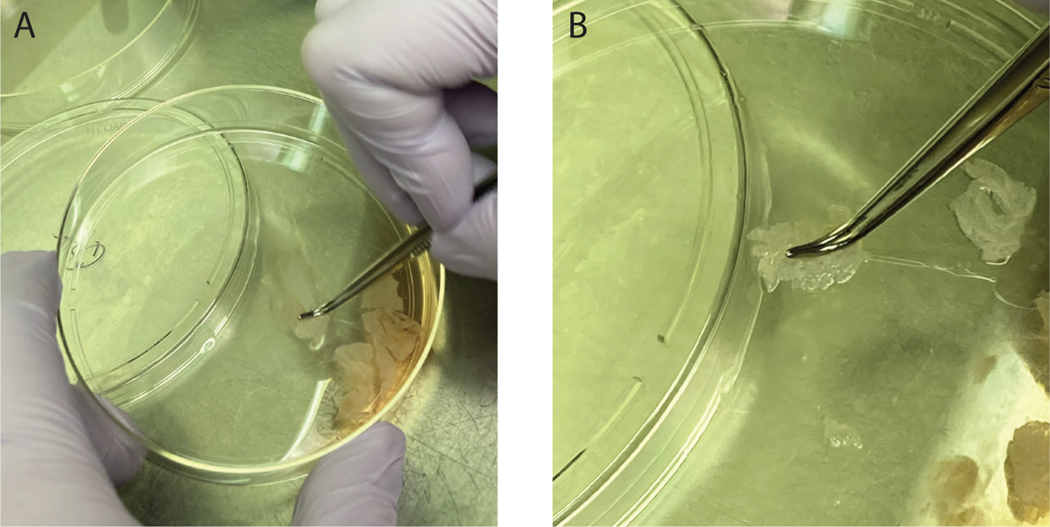
Figure 2.
NHEK and melanocyte isolation protocol. When scraping the tissue along the bottom of the dish to dislodge cells from the epidermal sheet it is easiest to use curved forceps to hold the tissue flat on the bottom of the dish. Note in (A) that the dish is held at an angle to allow the solution to pool at the bottom of the dish allowing the epidermal sheets to be scraped on an area of the plate not covered in solution. (B) shows a zoom in of the tissue being held to the bottom of the plate using curved forceps.

