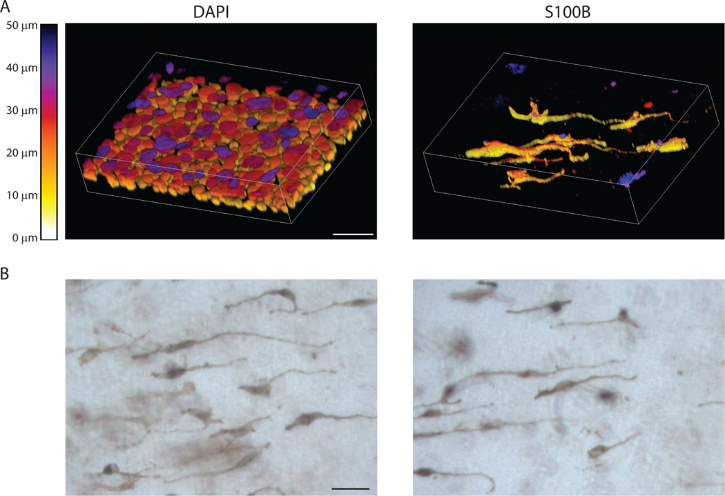
Figure 5.
Whole mount staining of organotypic cultures. A) Example images showing whole mount stained organotypic cultures stained with DAPI and S100B to label melanocytes. Images were collected using a Nikon A1R confocal laser scanning microscope equipped with GaAsP detectors and 20x Plan-Apochromat objective with a NA of 0.75. NIS-Elements was used to generate 3D images of z stacks using the alpha-blend method with z-depth color coding. Lookup table represents z-depth based on voxel color, scale bar = 50μm. B) Example images of whole mount samples taken with a brightfield microscope. These samples were not stained allowing visualization of the pigmented melanocytes and any pigment released into the culture. Scale bar = 50μm

