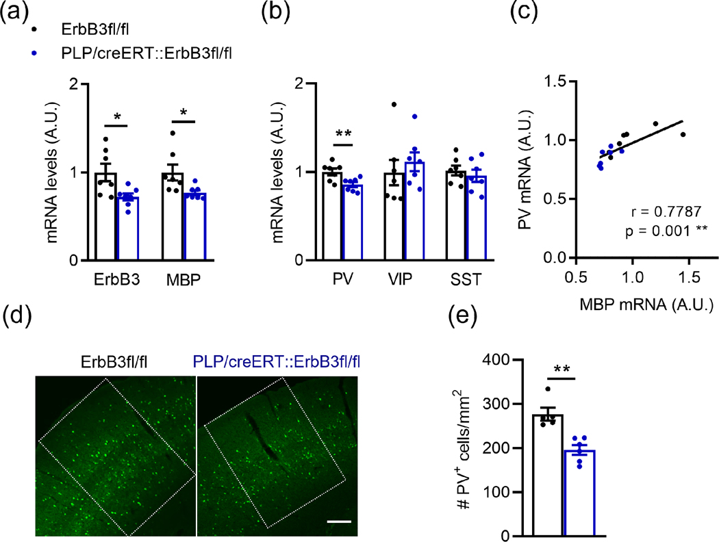
Figure 12: Loss of oligodendrocyte ErbB signaling by ErbB3 KO leads to reduced A1 MBP and PV mRNA levels and PV+ cell density.
a, Reduced ErbB3 (n = 7; p = 0.0230) and MBP (n = 7; p = 0.0175) mRNA levels in A1 after oligodendrocyte-specific ErbB3 KO (PLP/creERT::ErbB3fl/fl).
b, Relative mRNA expression for parvalbumin (PV), vasoactive intestinal peptide (VIP) and somatostatin (SST) in the A1 of WT (black) and PLP/creERT::ErbB3fl/fl (blue) mice. PV mRNA levels are reduced in A1 of PLP/creERT::ErbB3fl/fl mice (n = 6 – 7; p = 0.0093). No changes are observed in VIP (p = 0.3176) and STT (p = 0.62) mRNA expression.
c, A1 PV mRNA levels correlate with MBP mRNA levels (WT: black dots, PLP/creERT::ErbB3fl/fl: blue dots; Pearson’s correlation r = 0.7787; p = 0.001).
d, Representative photomicrographs of A1 sections from WT and PLP/creERT::ErbB3fl/fl mice showing PV+ cells (green). Scale bar: 100μm.
e, A1 PV+ cell density is reduced in PLP/creERT::ErbB3fl/fl mice (n = 5 – 6; p = 0.0043).
Mann-Whitney test was performed. Data are expressed as mean ± SEM.

