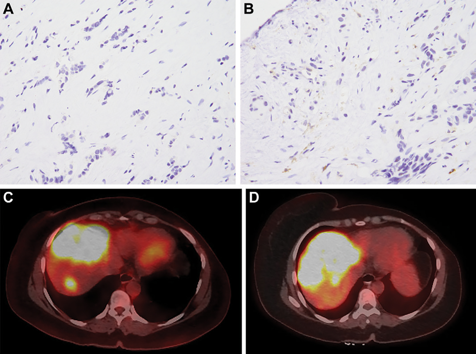
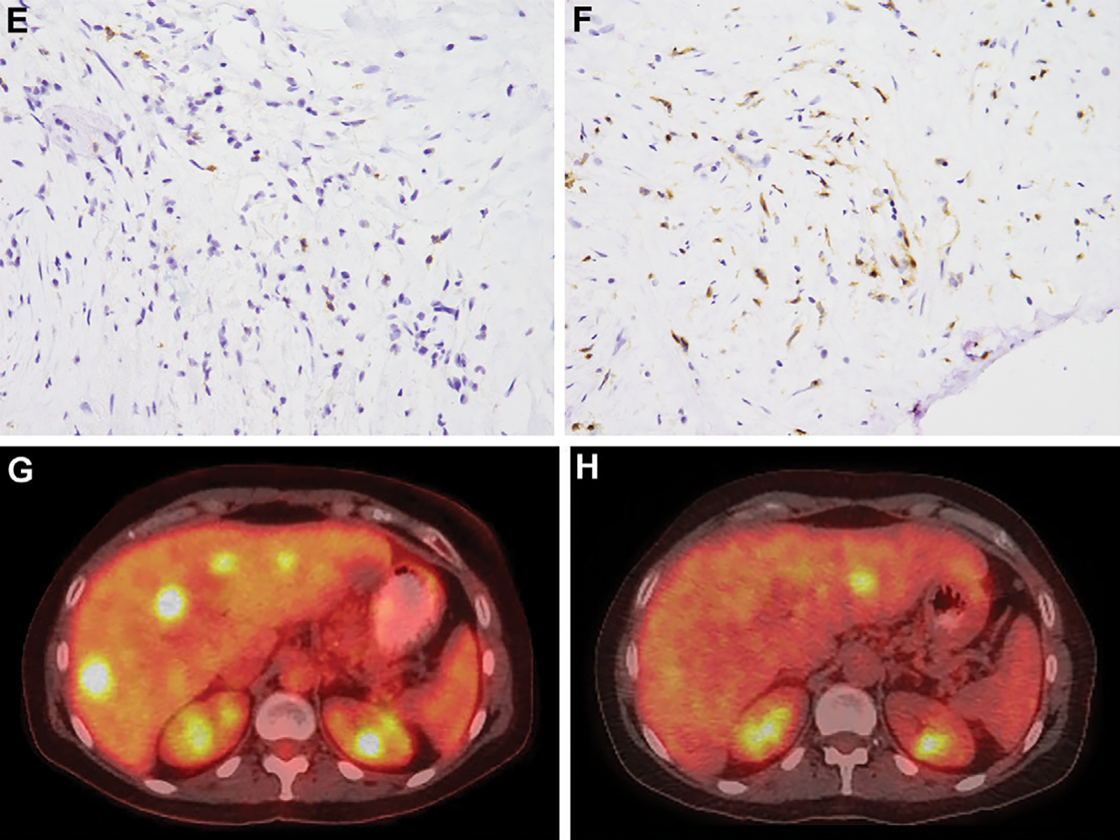
Figure 3:


A 47-year-old woman with metastatic breast cancer underwent transarterial radioembolization (TARE), with the first treatment to the right lobe of the liver. Pre-TARE baseline biopsy specimens show (A) low programmed cell death protein (PD) 1 staining (DAB brown) and (B) low CD4 staining (brown) (magnification, ×400). PET/CT images (C) before and (D) after TARE demonstrate progressive disease. A 51-year-old woman with metastatic breast cancer underwent TARE, with the first treatment to the right lobe of the liver. Pre-TARE baseline biopsy specimens show (E) high PD-1 staining and (F) high CD4 staining (magnification, ×400). PET/CT images (G) before and (H) after TARE demonstrate complete response.
