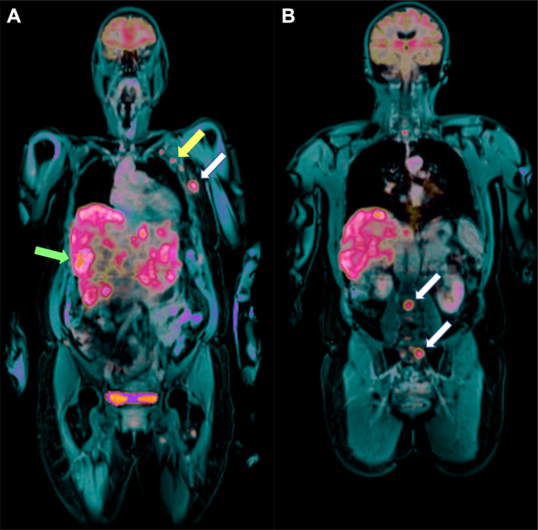
Figure 5.

(A and B) Fused PET and post-contrast fat-saturated T1-weighted imaging on the coronal plane (whole-body examination) shows liver and axillary involvement (green and yellow arrows in A, respectively) as well as rib and lumbo-sacral bone metastases (white arrows in A and B, respectively in in a 66-year-old patient with invasive ductal breast cancer (G3, ER/PgR−, HER2+) in the left breast (same patient as the patient in Figures 3 and 4).
