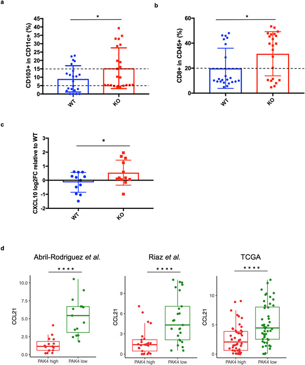
Figure 1: PAK4 expression levels are negatively associated with the presence of CD103+ dendritic cells in vivo and the levels of CCR7 ligand, CCL21, in biopsies of patients with melanoma.
a, Differences in the infiltration of CD103+ dendritic cells in B16 WT CC and PAK4 KO tumors (n = 44, 22 per group). Tumors were collected on day 6, after one dose of anti-PD-1. After processing and staining, CD103+ DCs were gated for singlets, live cells, CD45+, MHC-II+, CD11c+ and CD103+ cells. B16 PAK4 KO tumors had significantly higher levels of CD103+ DCs compared to B16 WT CC tumors (P = 0.04). b, Differences in the infiltration of CD8+ cells. Samples were gated for singlets, live cells, CD45+ and CD8+ population to have an estimate of the number of CD8 cells. B16 PAK4 KO samples showed a significant increase of CD45+/CD8+ cells compared to WT CC tumors (P = 0.02). c, RNA from a total of 24 in vivo samples (n = 12 per each group) were collected to perform RT-PCR. The cycle threshold (Ct) of each sample was normalized by the mean of the WT isotype group. CXCL10 expression is significantly increased in the PAK4 KO group. d, Differences in CCL21 log2FPKM expression levels between high and low PAK4 in biopsies of patients with melanoma (based on the upper and lower quartile) across three different clinical datasets: Abril-Rodriguez et. al., Riaz et. al., and TCGA. In all 3 cohorts, CCL21 levels were significantly enriched (P < 0.0001) in patients with low PAK4 expression. Statistical significance for Figure 1D was calculated using a two-tailed un-paired t-test. ****P < 0.0001.

