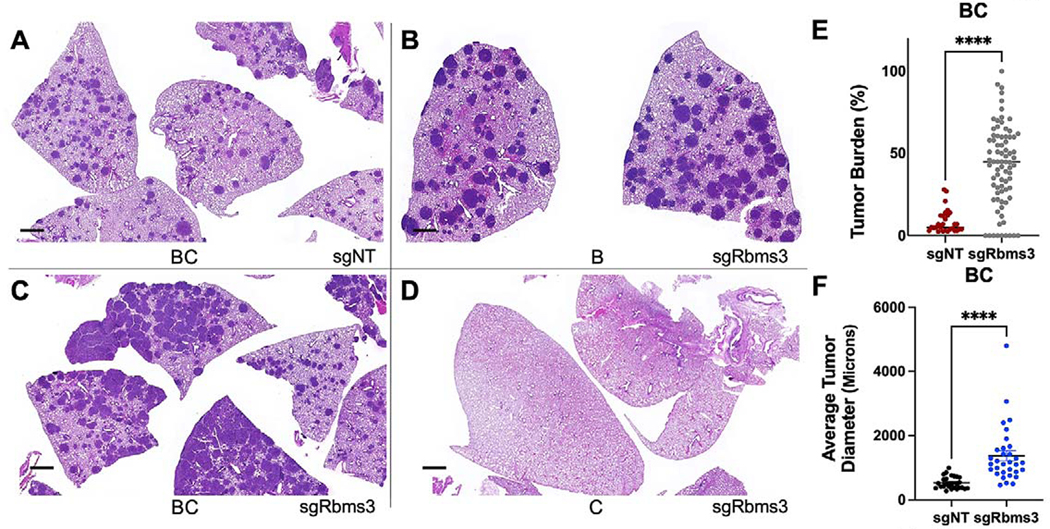
Figure 3. CRISPR/Cas9 editing of Rbms3 cooperates with BRAFV600E in a mouse model of lung cancer.
A-D: Representative images of different genotypes of harvested mouse lung sections following necropsy analyses stained with hematoxylin and eosin (H&E) 13 weeks post initiation with 5 × 10⁴ pfu lenti-CRE. CRISPR/CAS9-mediated genome editing was used in panels A, C, D to edit Rbms3 in vivo. Genotype and average tumor burden calculation of each experimental group was: A: sgNT-CRE virus in BrafCAT/+; H11LSL-CAS9 (BC) mice: 8.5%. B: sgRbms3-CRE virus in BrafCAT/+ (B) mice: 7.7%. C: sgRbms3-CRE virus in BC mice: 38.8%. D: sgRbms3-CRE virus in H11LSL-CAS9/+ (C) mice: 0%. Black bar in bottom left of each panel represents a 1000-micron scale bar. E: Quantification of individual tumor burden from genotypes in panel A compared to panel C. Tumor bearing lungs from panel B were identical to panel A. A paired T-test was used to determine statistical significance; p < 0.01.F: Quantification of tumor diameter was performed in microns using 25 individual tumors from genotypes in panel A compared to panel C using the 3D Histech MIDI Slide Scanner QuantCenter. Comprehensive analyses was conducted with over 200 lung tumors. N=50 mice individual or (biological replicates). N = 2 experimental replicates were performed comparing the indicated genotypes in A and C. Individual values are graphed, the black bar represents the mean, and the error bars represent SEM. A paired T-test was used to determine statistical significance; p < 0.0001.

