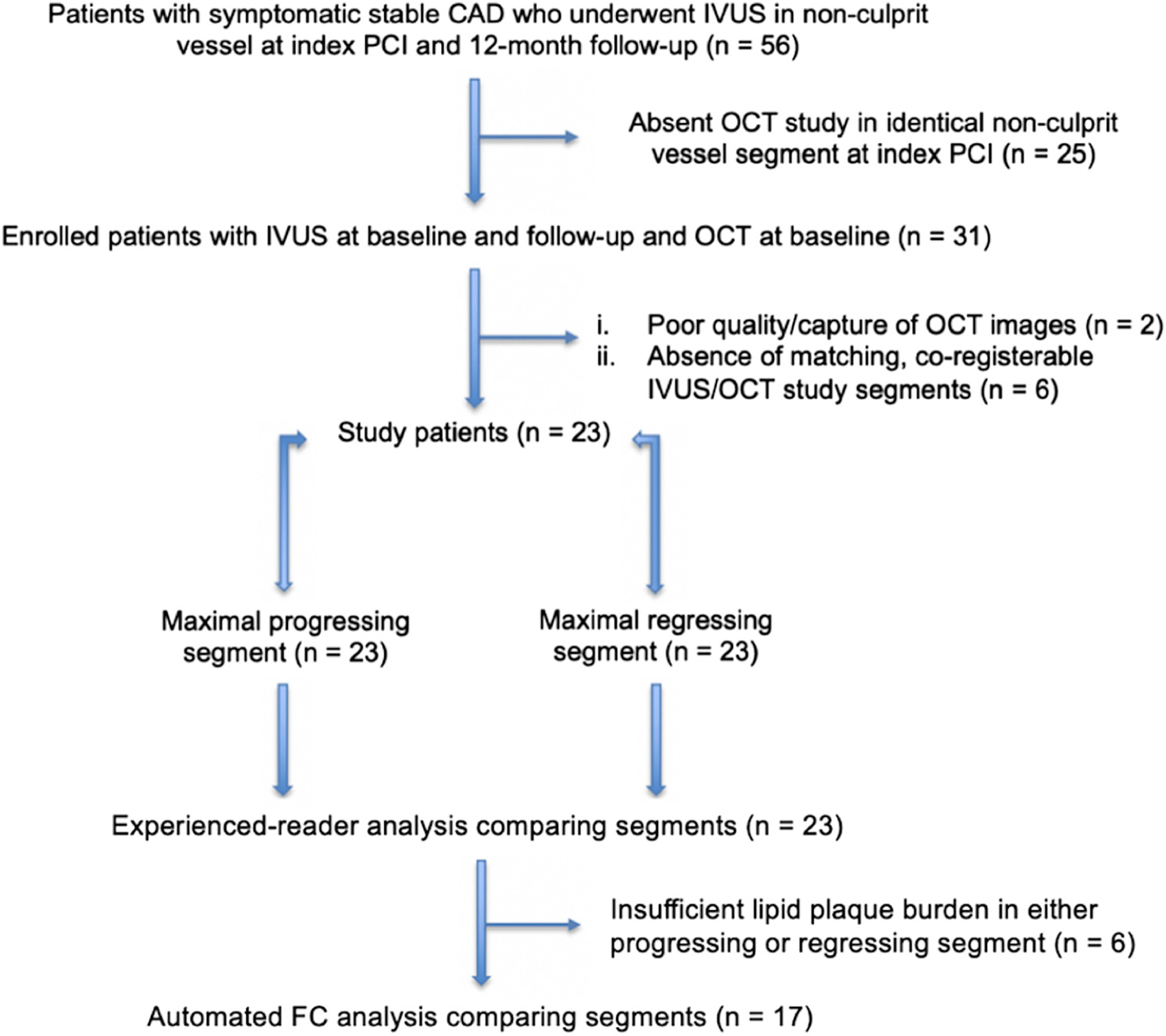
Figure. 1. Flowchart of study patient selection.

A total of 23 subjects with stable coronary artery disease who underwent nonculprit vessel imaging with optical coherence tomography and intravascular ultrasound at baseline and intrvascular ultrasound at follow-up were included in the study. Of these, 6 patients did not undergo fibrous cap analysis due to insufficient lipid plaque to assess these parameters. CAD, coronary artery disease; FC, fibrous cap; IVUS, intravascular ultrasound; OCT, optical coherence tomography; PCI, percutaneous coronary intervention.
