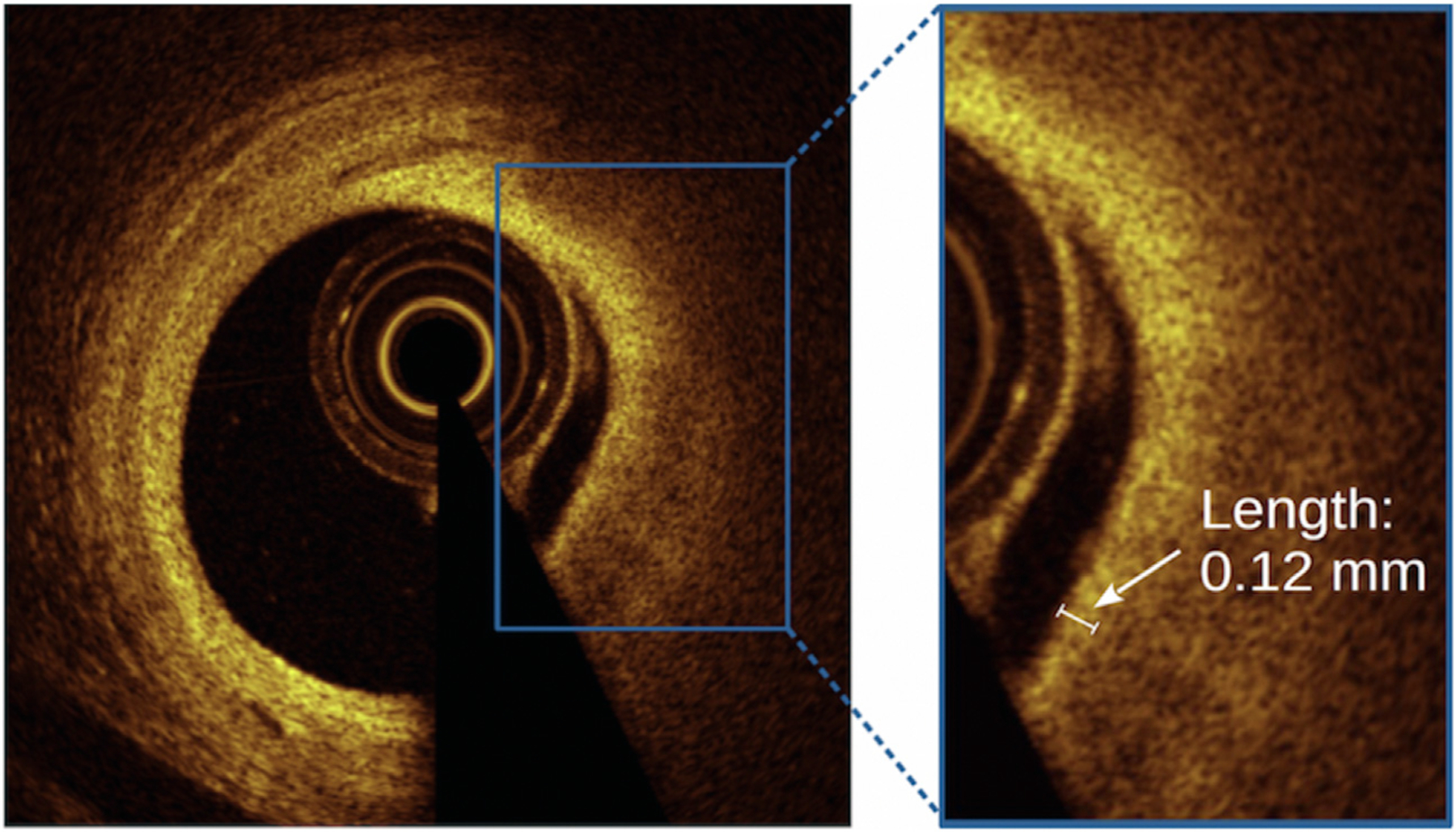
Figure. 2. Reader measurement of fibrous cap thickness.

Cross-sectional OCT images of signal-rich fibrous cap overlying a signal-poor lipid pool. The inset demonstrates the measurement of fibrous cap thickness (white arrow) within a frame, achieved using a length measurement tool on an offline OCT workstation. OCT, optical coherence tomography.
