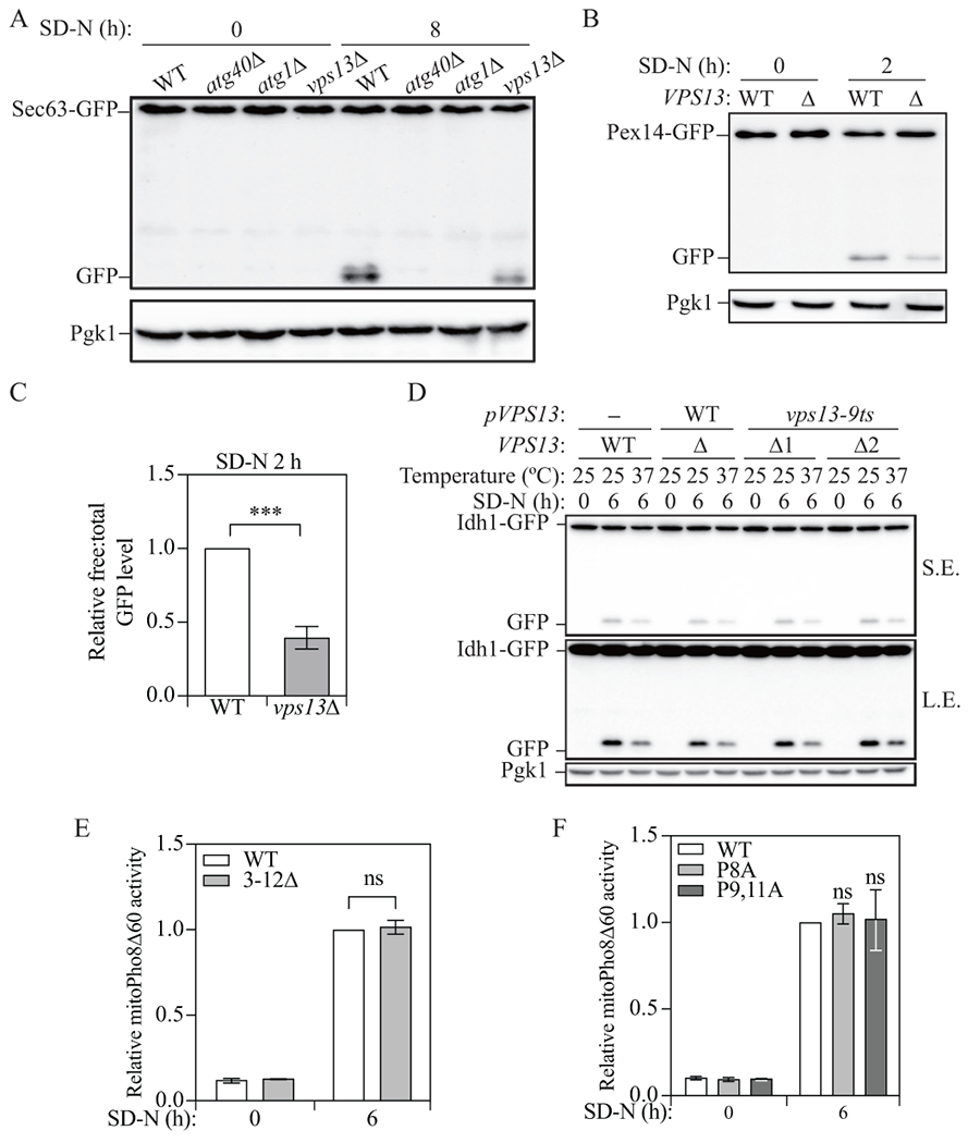
Figure 3.

Vps13 is important for reticulophagy and pexophagy, but not for mitophagy. (A) Protein samples were collected from wild-type (WXY233, Sec63-GFP), atg40Δ (DGY147, Sec63-GFP), atg1Δ (WXY283, Sec63-GFP), and vps13Δ (WXY234, Sec63-GFP) cells that were grown in YPD to mid-log phase (SD-N 0 h) and 8 h after nitrogen starvation. Western blots were probed with anti-YFP and anti-Pgk1 antibodies or antisera. (B) Wild-type (WXY201, Pex14-GFP) and vps13Δ (WXY203, Pex14-GFP) strains were cultured as indicated in Materials and Methods to induce pexophagy. The protein samples were collected from the culture in YTO (SD-N 0 h) and 2 h after nitrogen starvation (SD-N). Western blots were probed with anti-YFP and anti-Pgk1 antibodies or antisera. (C) Quantitative analysis of the relative free GFP to total GFP (Pex14-GFP + free GFP) level. The ratio of wild-type cells after 2 h starvation was set to 1 and other samples were normalized accordingly. (D) Wild-type cells with an empty pRS313 vector (YLY205) and vps13Δ cells with a pRS313 vector bearing wild-type Vps13 (YLY209) or the temperature-sensitive mutant Vps13 (YLY211, two colonies were included) were cultured as indicated in Materials and Methods to induce mitophagy. Protein samples were collected from the culture in SML-His (SD-N 0 h) at 25°C and 6 h after nitrogen starvation (SD-N) at either 25°C or 37°C. Western blots were probed with anti-YFP and anti-Pgk1 antibodies or antisera. (E and F) Cells expressing wild-type Mcp1 (YLY110, Mcp1-PA) and cells with truncated Mcp1 (YLY111) or single mutant (YLY115) and double mutant Mcp1 (YLY116) were cultured as indicated in Materials and Methods to induce mitophagy. Protein samples were collected from the culture in YPL (SD-N 0 h) and 6 h after nitrogen starvation. Mitophagy activity was monitored with the mitoPho8Δ60 assay. The mitoPho8Δ60 activities were normalized to wild-type cells after 6 h starvation. The error bar represents the SD of three independent experiments. Two-tailed t test was used for statistical significance. ***p < 0.001, ns, not significant.
