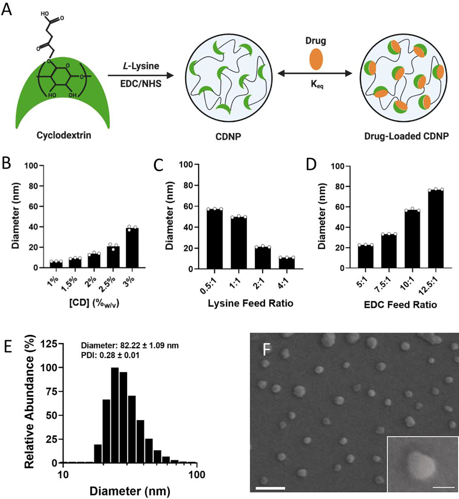
Figure 1. Development and characterization of CDNPs.

A) Schematic of cyclodextrin nanoparticle (CDNP) preparation through EDC/NHS-mediated crosslinking of succinyl-β-cyclodextrin by L-lysine with subsequent drug loading by guest-host interaction. CDNP diameter dependence on CD concentration (B; 10:1 EDC, 0.5:1 lysine), the molar ratio of lysine to succinyl groups (C; 3.3%w/v CD, 10:1 EDC), and the molar ratio of EDC to succinyl groups (D; 3.3%w/v CD, 0.5:1 lysine). E-F) CDNP characterization (3.3%w/v CD, 0.5:1 lysine, and 12.5:1 EDC). E) Number average histogram of particle size. Inset values: z-average diameter and polydispersity index (PDI); mean ± SD. F) Corresponding representative scanning electron microscopy images. Scale bar: 200 nm. Inset: higher resolution image of a single CDNP. Scale bar: 50 nm.
