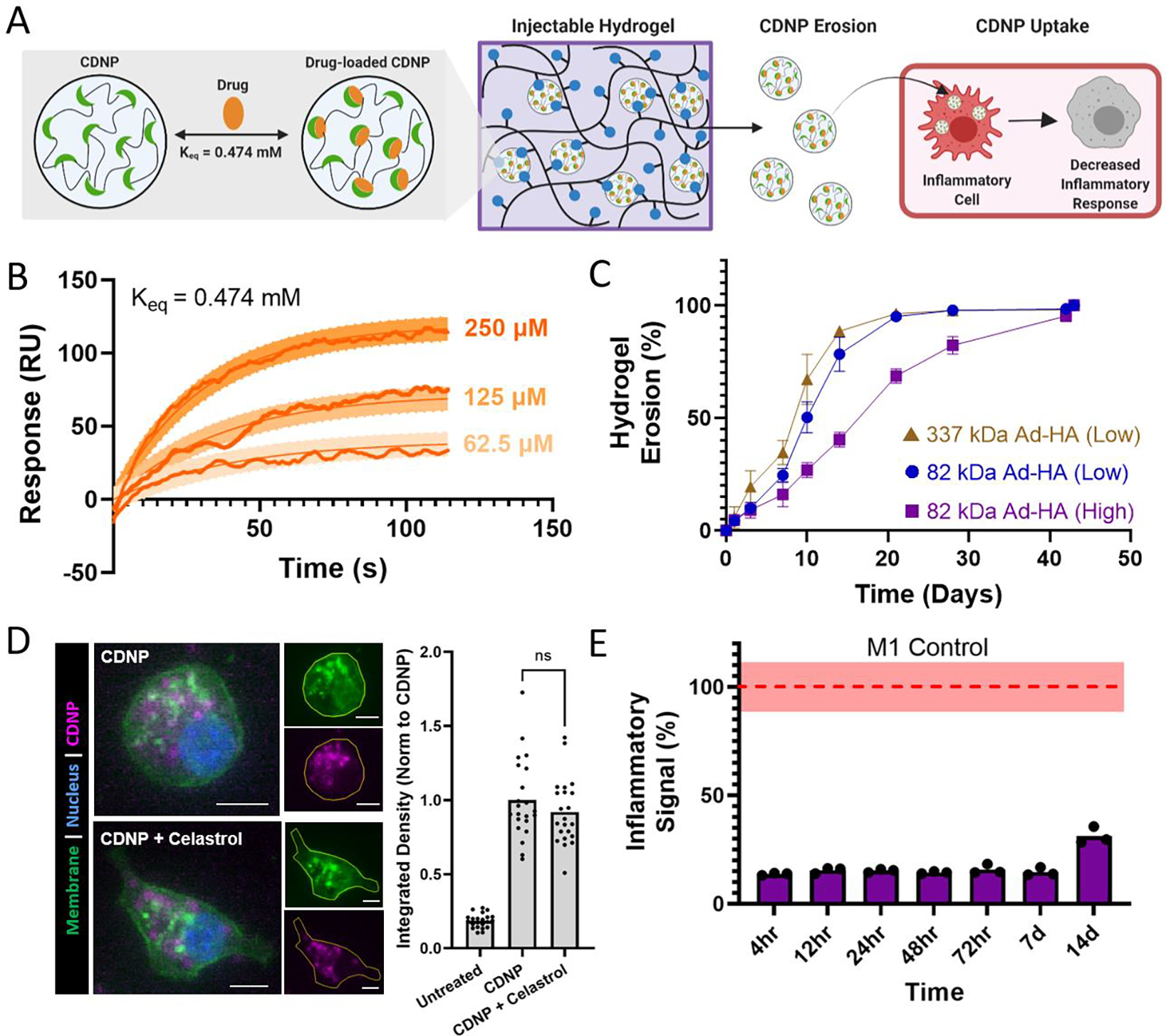
Figure 7. Therapeutic nanoparticle erosion enables long-term modulation of MF phenotype.

A) Schematic of drug loaded CDNP release from shear-thinning hydrogels and uptake by MF for desired decrease in inflammatory response. B) Binding sensograms between celastrol and CD, assessed at increasing concentrations of celastrol. C) Cumulative erosion for hydrogels formed from 337 kDa Ad-HA (Low, brown), 82 kDa Ad-HA (Low, blue), and 82 kDa Ad-HA (High, purple); mean ± SD, n = 4. D) RAW264.7 cell uptake of unloaded (CDNP) and drug-loaded (CDNP-Cel) nanoparticles from media conditioned by 82 kDa Ad-HA (High) hydrogel erosion. Representative images (left) show punctate accumulation of CDNP-AF555. Scale bars, 10 μm. Quantification of fluorescence per cell (right), normalized to unloaded CDNP. E) Anti-inflammatory activity of drug release samples, performed in RAW Blue™ cells by concurrent zymosan stimulation (100 μg/mL) and treatment with conditioned media from 82 kDa Ad-HA (High) hydrogels; p value < 0.0001 for all samples relative to zymosan-treated controls (dashed line, red). Data represent the mean ± SD, n = 3.
