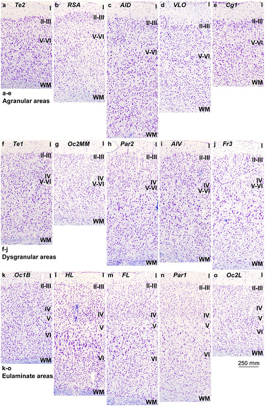
Fig. 2.
Cortical types across the rat cerebral neocortex. a–e, Micrographs of agranular mesocortical areas (Nissl staining). f–j, Micrographs of dysgranular mesocortical areas (Nissl staining). k–o, Micrographs of eulaminate areas (Nissl staining). Cortical areas are indicated according to Zilles (1985); see Table 4 for abbreviations of areas. Roman numerals indicate cortical layers. WM: white matter. Calibration bar in o applies to a–o

