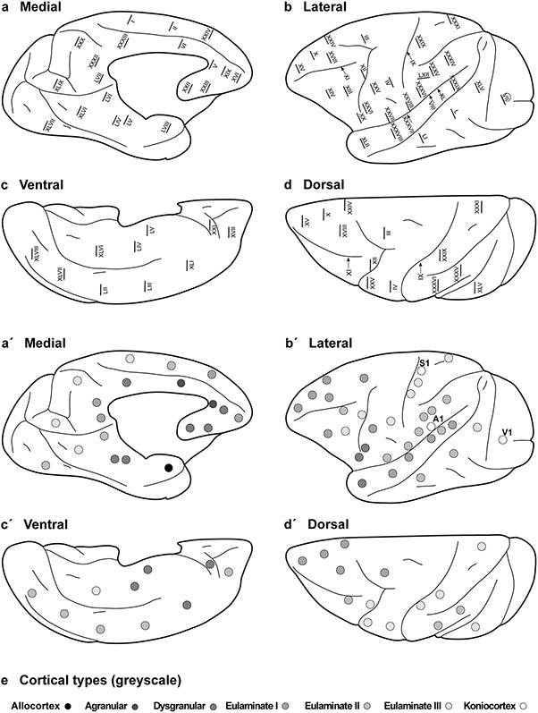
Fig. 4.
Distribution of cortical types across the Rhesus macaque cortex. Medial (a), lateral (b), ventral (c), and dorsal (d) views of the Rhesus macaque cerebral cortex; Roman numerals indicate the location of plates in the Atlas of von Bonin and Bailey (1947). a′–d′, Medial, lateral, ventral, and dorsal views of the Rhesus macaque cerebral cortex; dots in grayscale indicate cortical types for cortical areas photographed in the plates of the Atlas of von Bonin and Bailey (1947). Allocortical areas are colored in black; agranular mesocortical areas are colored with the darkest gray; dysgranular mesocortical and eulaminate areas are colored in progressively lighter grays. Koniocortical areas are colored in white. e, Grayscale of cortical types in a′–d′

