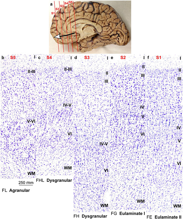
Fig. 5.
Cortical types across the human ventromedial prefrontal cortex. a, View of the left brain hemisphere (case HCD); red lines indicate planes of separation of coronal slabs; black and white arrow indicates gradient of laminar differentiation in the ventromedial prefrontal cortex (VMPFC). b–f, Micrographs of areas across the VMPFC (Nissl staining) at the levels indicated in A. Cortical areas, according to von Economo and Koskinas (1925/2008), and cortical types, according to laminar features of Table 3, are indicated below each micrograph. S1, S2, S3, S4, S5, indicate coronal slabs from anterior to posterior. Roman numerals indicate cortical layers. WM: white matter. Calibration bar in b applies to b–f

