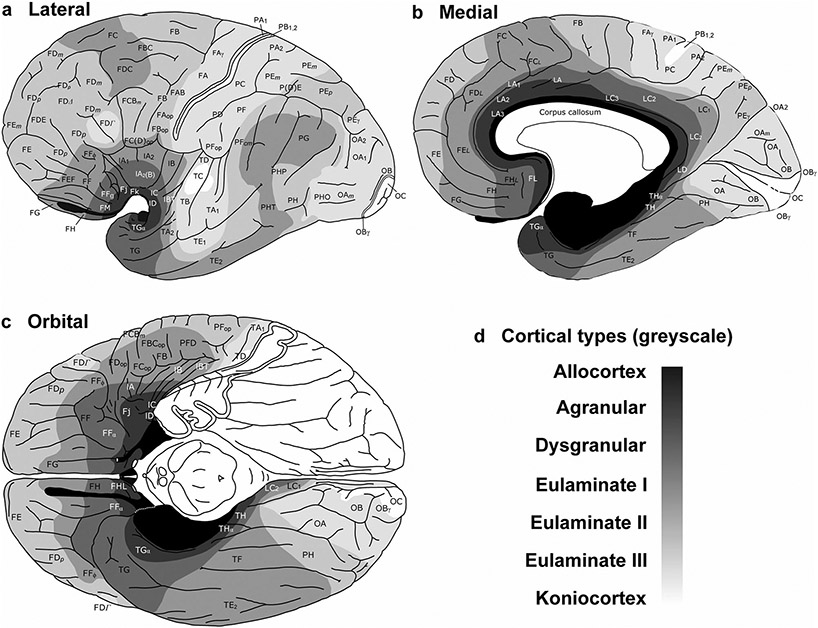
Fig. 6.
Distribution of cortical types across the human cortex. Lateral (a), medial (b), and orbital (c), views of the human brain. Cortical areas are indicated according to von Economo and Koskinas (1925/2008); cortical types are colored in grayscale according to García-Cabezas et al. (2020). Allocortical areas are colored in black; agranular mesocortical areas are colored with the darkest gray; dysgranular mesocortical and eulaminate areas are colored in progressively lighter grays. Koniocortical areas are colored in white. d, Grayscale of cortical types in a–c. This figure is modified from a previous article of our group [see Fig. 8 from García-Cabezas et al. (2020)] published under Creative Commons CC-BY license that grants to third parties all content of the article (https://www.frontiersin.org/legal/copyright-statement)

