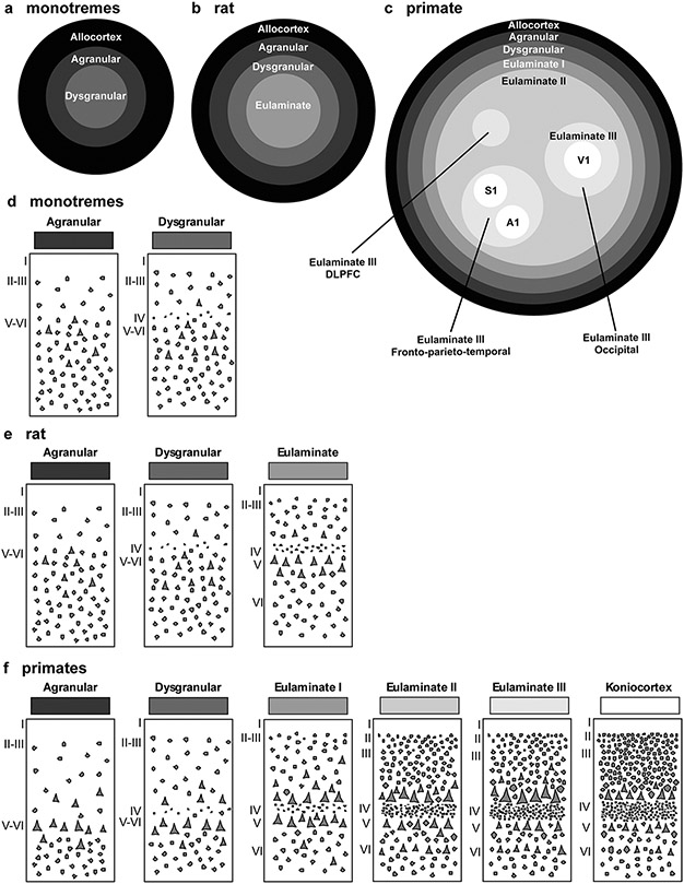
Fig. 7.
Distribution of cortical types in simplified flat maps of monotremes, rats, and primates. a–c, Simplified flat maps of the cerebral cortex of monotremes (a), rats (b), and primates (c). Cortical types are colored in grayscale; allocortical areas are colored in black; agranular mesocortical areas are colored with the darkest gray; dysgranular mesocortical and eulaminate areas are colored in progressively lighter grays. Koniocortical areas are colored in white. d, Cartoons of types of neocortical areas in monotremes. e, Cartoons of types of neocortical areas in rats. f, Cartoons of types of neocortical areas in primates. Roman numerals indicate cortical layers. A1 primary auditory area, DLPFC dorsolateral prefrontal cortex, S1 primary somesthetic area, V1 primary visual area

