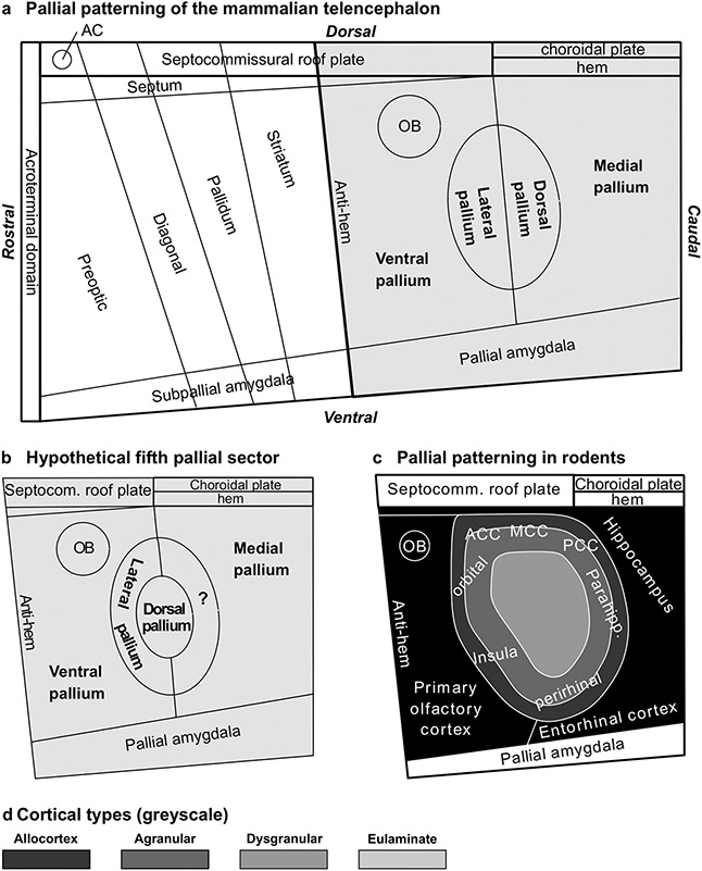
Fig. 8.
Pallial patterning in mammals. a, Sketch of Fundamental Morphological Units (FMUs) of the mammalian telencephalon according to the Prosomeric Model (Puelles and Rubenstein 1993, 2015; Nieuwenhuys and Puelles 2016; Puelles 2018; Puelles et al. 2019). Gray shading indicates pallial FMUs according to the Tetrapartite Model of Pallial Development and Evolution (Puelles 2017, 2021). b, Sketch of FMUs of the mammalian pallium showing a hypothetical fifth pallial sector indicated with interrogation sign (?). c, Sketch of the rat pallium according to pattering studies in rodents (Puelles et al. 2019); prospective allocortical areas are colored in black; prospective agranular mesocortical areas are colored with the darkest gray; prospective dysgranular mesocortical and prospective eulaminate areas are colored in progressively lighter grays. d, Grayscale of cortical types in c. Abbreviations: AC, anterior commissure; ACC, anterior cingulate cortex; MCC, middle cingulate cortex; OB, olfactory bulb; PCC, posterior cingulate cortex

