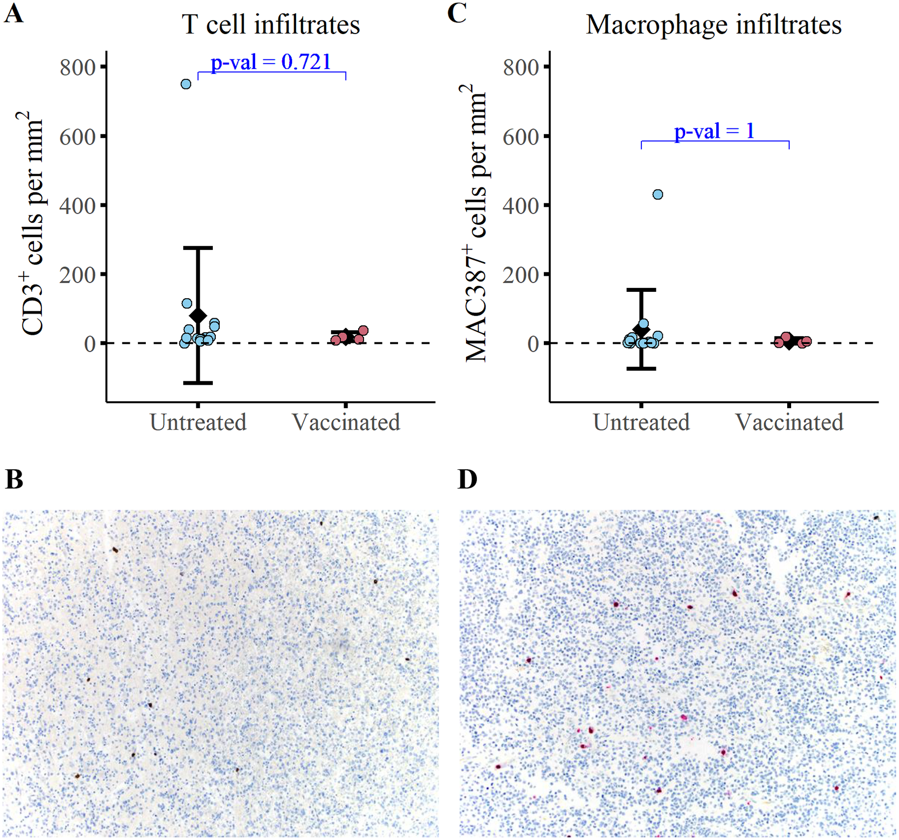
Figure 5. Immunized dogs exhibit no detectable change in tumor infiltrating immune cells relative to untreated controls at the time of necropsy.

Quantification of (A) T cell infiltrates (CD3+) and (C) macrophage infiltrates (MAC387+) at the time of necropsy in 4 study dogs and 14 untreated dogs with gliomas. Representative images of CD3 immunolabeling (B) and MAC387 immunolabeling (D) are depicted. A Wilcoxon two sample t-test was conducted to evaluate statistical significance.
