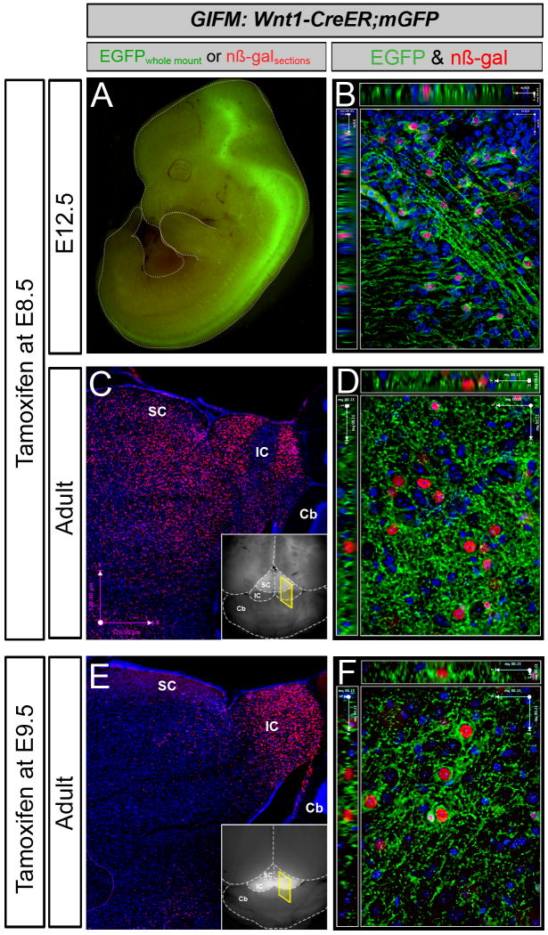
Figure 1. Representative Results for Wnt1-fate mapping.
Examples of whole-mount and immunocytochemistry results in Wnt1-CreERT;mGFP embryos and adult brains fate mapped (marked) at E8.5 (A-D) and E9.5 (E-F). A. Wnt1-CreER;mGFP embryos at E12.5 exhibit GFP fluorescence primarily in the midbrain (Mb), posterior hindbrain (Hb) and spinal cord (Sp). B. Marked cells visualized and analyzed at the cellular level by immunohistochemistry as shown on this 1-μm thick optical section (40× objective, z-series acquisition). Nuclear β-galactosidase (nβ-gal) antibody labeling (red) and GFP antibody labeling (green) indicates fate-mapped cells shown in the ventral midbrain. Here, recombined cells are double positive (nβ-gal+/GFP+) because of the nature of the conditional mGFP reporter. C. Whole-mount florescence microscopy reveals faint GFP fluorescence in the superior colliculi (SC) of the adult (inset). Assessing the Wnt1 lineage marked at E8.5 on adult sections with low magnification (5×) microscopy reveals that the Wnt1 lineage gives rise to midbrain structures including the superior (SC) and inferior (IC) colliculi (C). D. A 40×, 1 mm optical section taken from the ventral midbrain in the vicinity of dopaminergic neurons with nβ-gal+ cells and a rich GFP+ axonal plexus. E. Marking at E9.5 results in substantial GFP labeling that is readily observed in the inferior colliculus of the midbrain by whole mount fluorescence (inset). The Wnt1 lineage marked at E9.5 on adult sections (β-gal+, red) is concentrated in the inferior colliculus (dorsal-posterior midbrain) as seen with low magnification (5×) microscopy (E). F. A 40×, 1 mm optical section taken from the ventral midbrain in the vicinity of dopaminergic neurons with nβ-gal+ cells and a rich GFP+ axonal plexus. Wnt1-derived neurons marked at E8.5 versus E9.5 are progressively restricted from contributing to dorsal midbrain, while the Wnt1 lineage persists in contributing to ventral midbrain.

