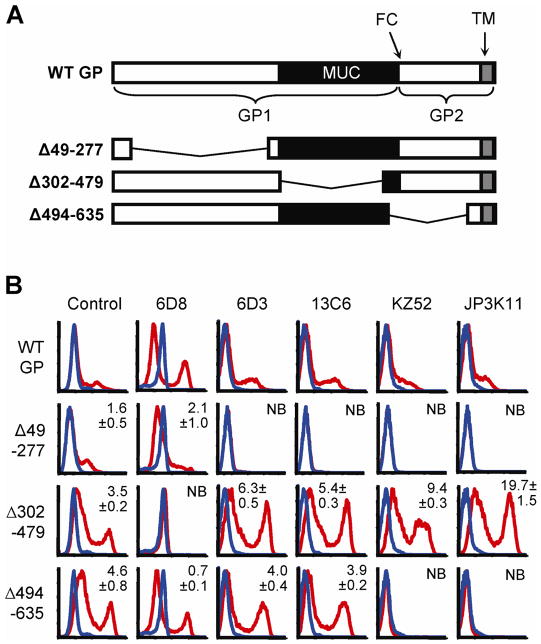
Figure 2. N-terminal GP1 contains potent neutralization epitopes.
(A) Cartoon of WT GP and deletion mutants Δ49-277, Δ302-479, and Δ494-635; GP1 and GP2 subunits, furin cleavage site (FC), MUC (black) and transmembrane (TM; gray) are indicated. (B) FACS staining of GP deletion mutants expressed in 293 cells; GP expressing- (red lines) or empty vector-transfected (blue lines) cells were stained with control serum (sGP/GP), 6D3, 13C6, KZ52, or JP3K11. Experiments were performed at least four times with similar results and numbers represent binding ratios (MFI relative to WT) ± SEM.

