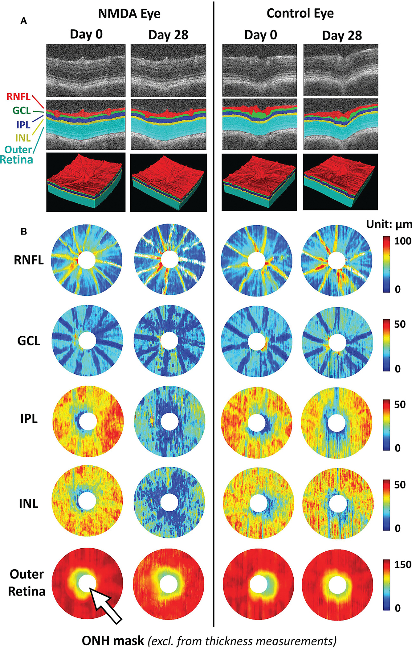
FIGURE 2.

Representative images of deep learning-assisted automatic retinal layer segmentation (A) and the thickness measurements of 5 retinal layers for both injured and control rat eyes (B) before and 28 days after unilateral N-methyl-D-aspartate (NMDA) injection. Automatic retinal layer segmentation was achieved using LF-UNet - an anatomical-aware cascaded deep-learning-based retinal optical coherence tomography (OCT) segmentation framework that has been validated on human retinal OCT data (42). In this work, two techniques were applied to improve the efficiency and generalizability of the LF-UNet segmentation framework when training with a small, labeled dataset – 1) composited transfer-learning and domain adaptation, and 2) pseudo-labeling. [excerpted from (42)]. (RNFL, retinal nerve fiber layer; GCL, ganglion cell layer; IPL, inner plexiform layer; INL, inner nuclear layer; ONH, optic nerve head).
