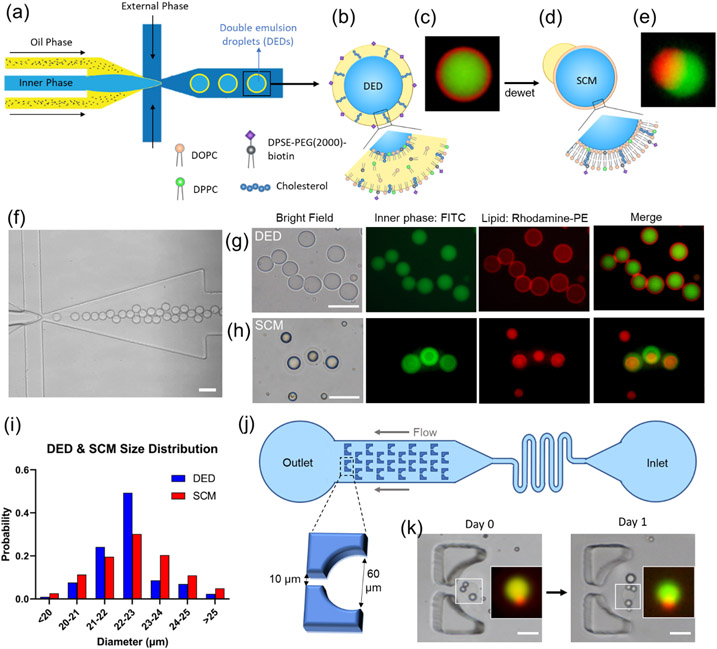
Figure 2.
Generation of double emulsion droplets (DEDs) and single-compartment multisomes (SCMs). (a) Illustration of DED generation on a flow-focusing microfluidic device. (b) Illustration figure and (c) fluorescence image of DED. (c) Images of DEDs generated with 0.05 mol% rhodamine-PE lipids (red) encapsulating FITC (green). (d) Illustration figure and (e) fluorescence image of SCMs converted from DEDs, showing SCMs have an oil cap and a lipid bilayer membrane. (f) Generation of DEDs. (g) Images of DEDs. (h) Images of SCMs. (i) Size distribution of DED and SCM. (j) Illustration of the trapping array. (k) SCMs are stable in the trapping array filled with RPMI + 10% FBS after one day. Scale bar = 50 μm. N = 302 for DEDs. N=265 for SMCs.

