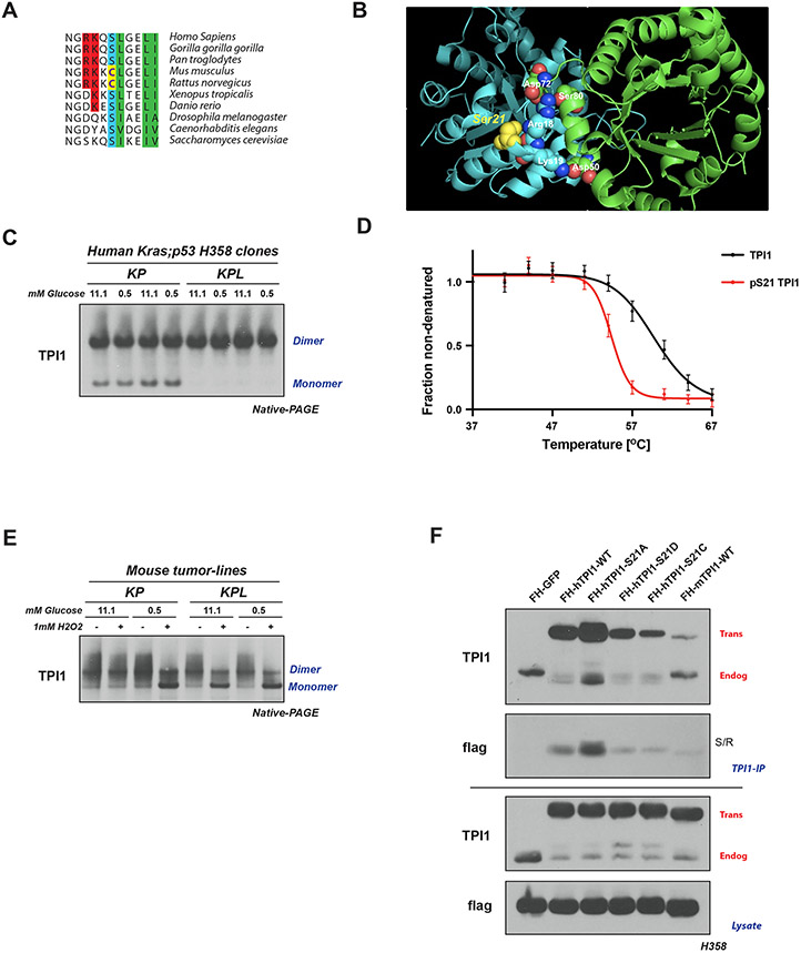
Figure 5. LKB1 regulates the multimeric state of hTPI1 but not mTpi1 due to an amino acid difference at position 21.
(A) Sequence alignment of TPI1 amino acid residues 16 to 26 across species, showing conservation of Ser21 from H. sapiens to S. Cerevisiae, with cysteine at position 21 in mouse and rat Tpi1. Cartoon comparing predicted side-chain chemistry, with oxidized cysteine and phosphorylated serine, is drawn below. (B) Crystal structure of TPI1 homodimer (cyan and green respectively) with critical residues highlighted in space-filling atoms. Serine 21 on the cyan monomer is highlighted in yellow. (C) Western blot analysis of Blue Native PAGE of human isogenic clones derived from KP H358 hLUAD cell-line. Cells were grown under normal (11.1 mM) or low (0.5 mM) glucose conditions for 6 hr prior to collection. (D) Melting curve plot from Thermal Profiling of unmodified and Serine 21 phosphorylated TPI1. Analysis conducted in H2009 and H358 isogenic clones expressing Cas9 and a non-targeting (sgNT1.1 and sgNT1.2 or sgNT1.4 and sgNT1.6, respectively) guide RNA. Data presented is from seven biological replicates and reported as the mean (−/+ S.D.) (E) Western blot (Blue Native PAGE) of extracts from mLUAD cell lines. Cells were cultured in either 11.1 mM or 0.5 mM glucose for 6 hours then treated with 1 mM H2O2 for 15 minutes. (F) Western blot of proteins co-immunoprecipitated from extracts of H358 cells expressing Cas9 and a non-targeting (FH-GFP cell line) or TPI1-specific (all other cell lines) guide RNA and transgenic expression of Flag-HA tagged GFP or guide RNA resistant TPI1 allelic variants using a polyclonal antibody against full-length TPI1. Cells were cultured in 0.5 mM glucose for 6 hr prior to collection.

