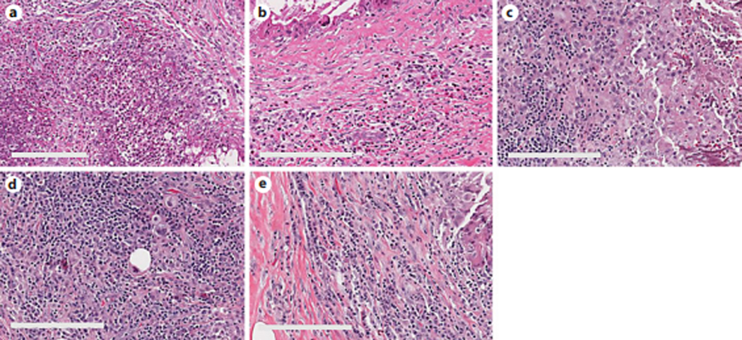
Fig. 2.

H&E images of inflammatory cells adjacent to the biopsy needle tract. a, b Neutrophils and eosinophils were present 31 days after the biopsy. c, d Macrophages and lymphocytes remained present 56 days after the biopsy. e Plasma cell-rich area adjacent to the needle tract 25 days after the biopsy. Scale bars, 50 μm.
