
Figure 4.
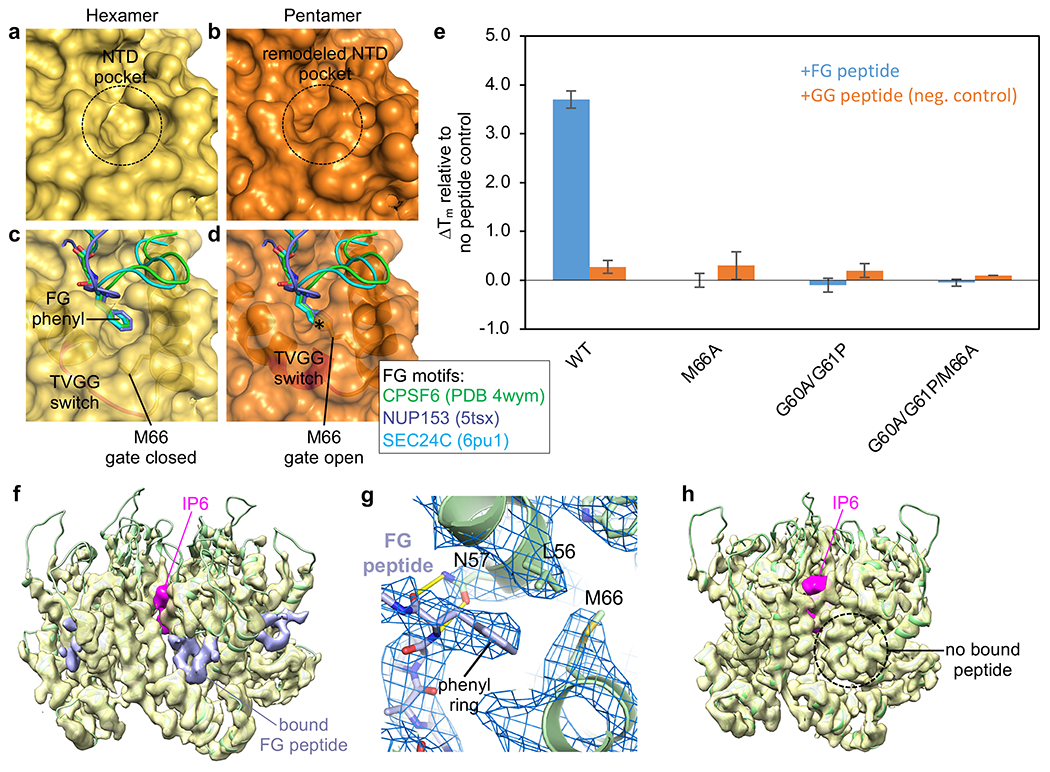
Conformational switching of the TVGG motif remodels the FG-motif binding site. (a,b) Connolly surface representations of our WT hexamer and pentamer structures. (c,d) The hexamer and pentamer are superimposed with the indicated crystal structures of CA with bound FG ligands. * indicates steric clash. (e) Thermostabilization of indicated CLPs by CPSF6-FG and CPSF6-GG peptides. (f,h) Views of the hexamer and pentamer from a cryoEM reconstruction (3.9 Å) of the declination from WT CLPs incubated with excess CPSF6-FG peptide (0.4 contour). Pink indicates bound IP6, found in both hexamer and pentamer. Lavender indicates that the peptide is only bound to the hexamer. (g) Close-up of the binding interaction in the hexamer pocket.