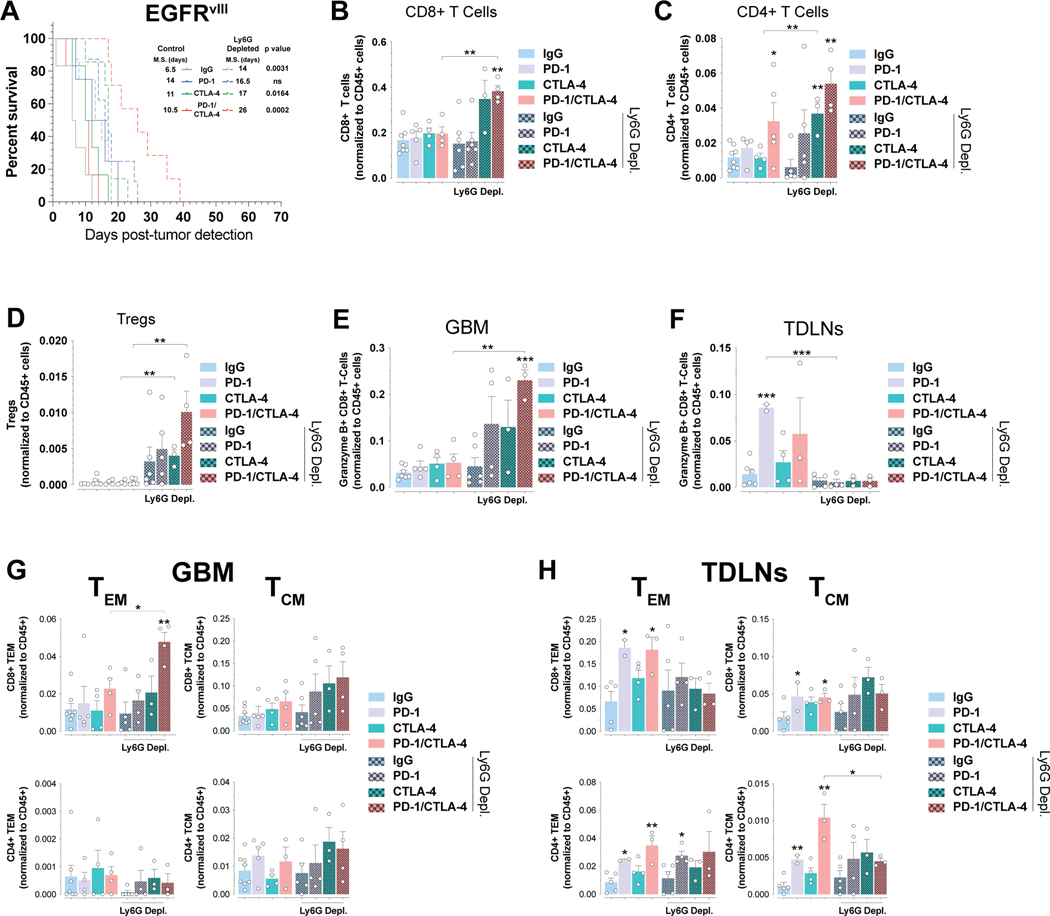
Figure 5. PMN-MDSCs depletion sensitizes EGFR-driven GBMs to PD-1/CTLA-4 checkpoint blockade treatments.
A) Kaplan-Meier analysis of EGFRvIII GBM mice treated as indicated. p values log-rank (Mantel Cox) test. B-D) Relative numbers of CD8+ (B) CD4+ (C) and (D) regulatory T cells. E-H) Relative numbers of Granzyme B+ CD8+ T cells in GBMs (E) and TDLNs (F) and CD8+ and CD4+ T cell subset from GBMs (G) and TDLNs (H) of PMN-MDSC (anti-Ly6G) depleted EGFRvIII mice treated as indicated. Non-bracketed comparisons to IgG controls (B-H). Mean±SEM of biological replicates. *p<0.05, **p<0.01, ***p<0.001, ****p<0.0001, unpaired t test, two-tailed.

