Fig. 6.

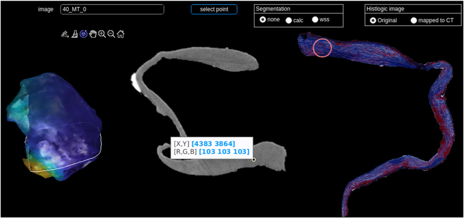
Visual exploration of combined micro-CT and histology; left: 3D model with WSS, the white line indicates the current slice; middle: segmented micro-CT image, right: segmented, corresponding MT-stained histologic image

Visual exploration of combined micro-CT and histology; left: 3D model with WSS, the white line indicates the current slice; middle: segmented micro-CT image, right: segmented, corresponding MT-stained histologic image