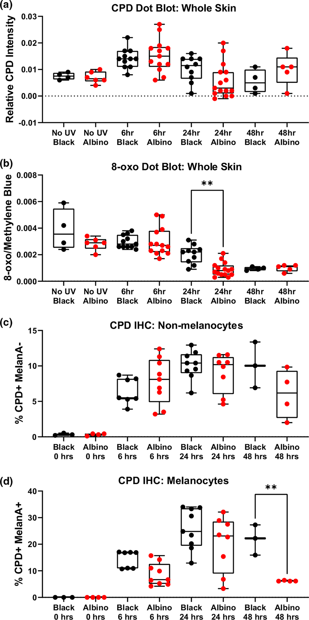
FIGURE 5.

Cell-intrinsic melanin does not protect melanocytes or keratinocytes from post-UV DNA damage. (a, b) Box and whisker plots show the amount of CPD (“a”) and 8-oxoG (“b”) lesions detected in dot blots of dorsal skin harvested from black or albino TpN mice prior to, and 6, 24, or 48 h after UVB irradiation. CPD and 8-oxoG signals were normalized to methylene blue staining. (c, d) Box and whisker plots showing the amount of immunofluorescent CPD staining in dorsal skin samples from black or albino TpN mice harvested prior to, 6, 24, or 48 h after UV irradiation. Co-staining with MelanA+ was used to identify keratinocytes (“c”; Melan-A−) and melanocytes (“d”; Melan-A+), respectively. (a–d) Each dot represents one biological sample. Boxes span the 25th–75th percentile with whiskers from the minimum to maximum value. Statistical significance was determined using ANOVA with a Dunnett’s T3 multiple comparisons test.
