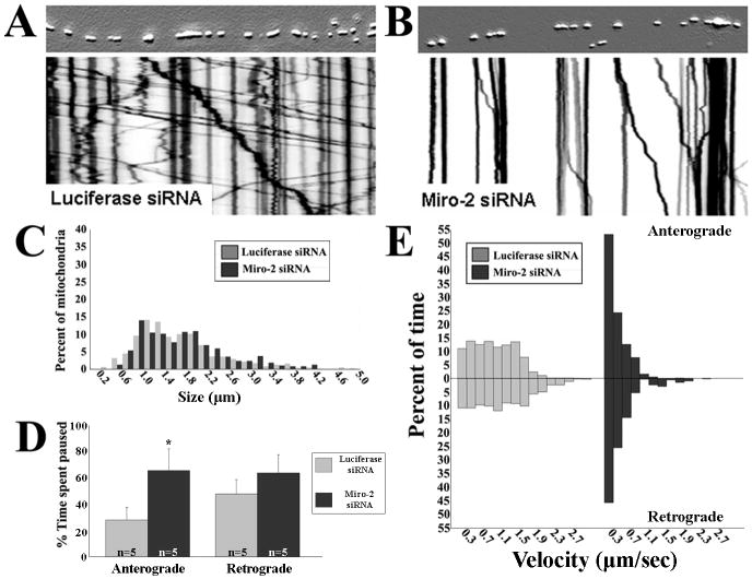
Figure 5. Depletion of Miro2 produces a mitochondria transport abnormality similar to that observed with loss of Mfn2 in DRG neurons.
(A, B) Kymograph analysis reveals that siRNA-mediated knockdown of Miro2 dramatically altered patterns of mitochondrial transport. (C) Size frequency histogram of axonal mitochondria demonstrated that depletion of Miro2 altered mitochondrial transport without changing mitochondrial morphology. (D) Similar to Mfn2-/- cultures, mitochondria spent a greater percentage of time paused between anterograde movements in Miro2 knockdown cultures (* = p<0.001, t-test; n = # of axons from which image stacks were created). Pauses between retrograde movements trended toward longer pause times but did not reach statistical significance. (E) Mitochondria velocity distributions were also skewed toward slower movements in Miro2 knockdown cultures, similar to effects seen with loss of Mfn2.

