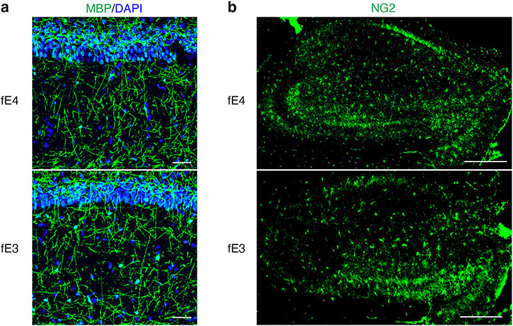
Extended Data Figure 3. No observable difference in MBP or NG2 immunostaining between fE4 and fE3 mice in the absence of human mutant tau.
a, Representative images of myelin sheath staining with anti-MBP and DAPI in the stratum radiatum underneath the pyramidal cell layer of CA1 of the hippocampus in 10-month-old fE4 and fE3 mice (scale bar, 50 μm). b, Representative images of oligodendrocyte progenitor cell (OPC) staining with anti-NG2 in the hippocampus of 10-month-old fE4 and fE3 mice (scale bar, 500 μm). Experiments depicted in representative images in a–b were performed on n=4 mice per genotype using 2 brain sections per mouse, with reproducible data.

