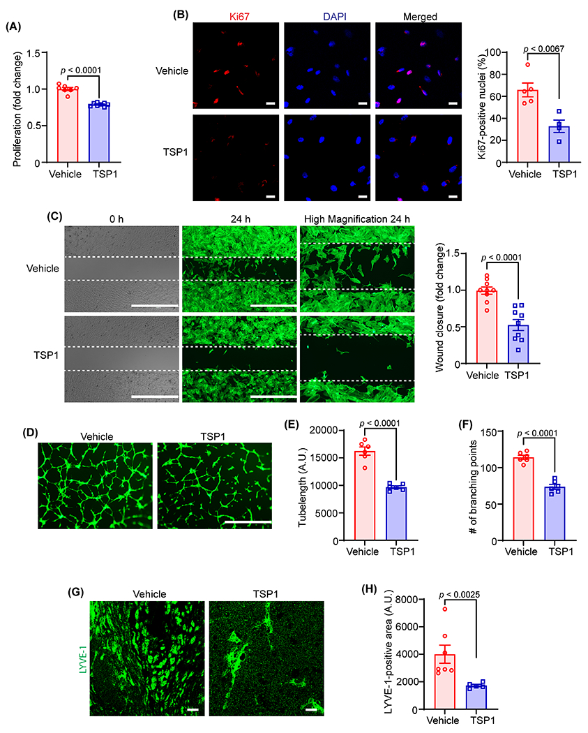
Fig. 2. TSP1 suppresses both in vitro and in vivo lymphangiogenesis.

(A) Vehicle- and TSP1-pretreated (22 nM, 4 h) human LEC were stimulated with VEGF-C (100 ng/mL) and proliferation investigated after 48 h using WST-1 assay. Data are representative of three independent experiments performed at least in duplicate. (B) LEC-plated on coverslips were pretreated and stimulated as in (A) for 24 h. Cells were immunostained for Ki67 (red) and nuclei counterstained with DAPI (blue). Images of at least four random microscopic fields were captured. Representative images are shown. Scale bar 20 μm. Bar graph represents the percentage of Ki67-positive nuclei (n = 4-5). (C) LEC migration in response to vehicle (VEGF-C) and TSP1 (VEGF-C+TSP1) was investigated after 24 h using Culture-Insert 2 Well 24 (ibidi USA). Representative images of wounds at 0 h and 24 h are shown. Scale bar 1000 μm. Bar diagram shows quantification of wound closure (n = 9). (D) Vehicle- or TSP1-pretreated LEC were seeded in wells of a Matrigel-coated plate in basal medium containing VEGF-C ± TSP1 and tube formation determined after 6 h. Representative images of tube formation are shown. Scale bar 1000 μm. Images of random fields were captured, and tube length (E) and number of branching points (F) quantified (n = 6). (G-H) Wild-type male mice were injected s.c. with Matrigel solutions premixed with either VEGF-C or VEGF-C+TSP1. Plugs were isolated after 10 days, sectioned and immunostained for LYVE-1. (G) Representative images of LYVE-1 staining of cross-sections of the Matrigel plugs are shown. Scale bar 20 μm. (H) Quantification of LYVE-1-positive area (n = 5-7). Statistical analyses were performed using a two-tailed unpaired student t-test (A-C, E and F) and a Mann-Whitney test (H). Data represent mean ± SEM. LYVE-1, lymphatic vessel endothelial hyaluronan receptor-1.
