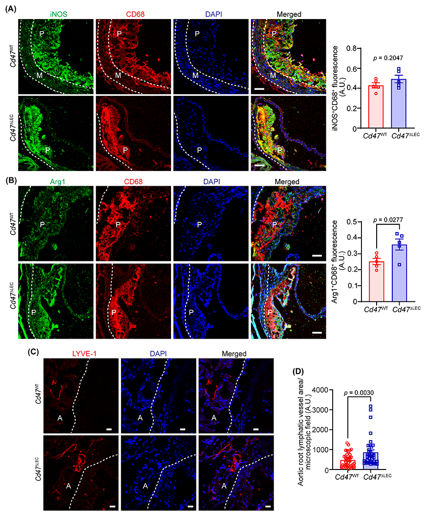
Fig. 6. LEC-specific deletion of Cd47 in mice increases arterial LV density.

(A-D) Aortic root cross-sections from male AAV8-PCSK9-injected Cd47WT and Cd47ΔLEC mice (16 weeks Western diet) were immunostained for CD68, iNOS, Arg1 and LYVE-1. Nuclei were counterstained with DAPI (blue). Representative confocal images of iNOS (green, A) and CD68 (red); Arg1 (green, B) and CD68 (red) Scale bar 50 μm; LYVE-1 staining (red, C) Scale bar 20 μm, are shown (n = 5-6). Statistical analyses were performed using a two-tailed unpaired student t-test (A and B) and a Mann-Whitney test (C). Data represent mean ± SEM. P: plaque, A: adventitia and M: media.
