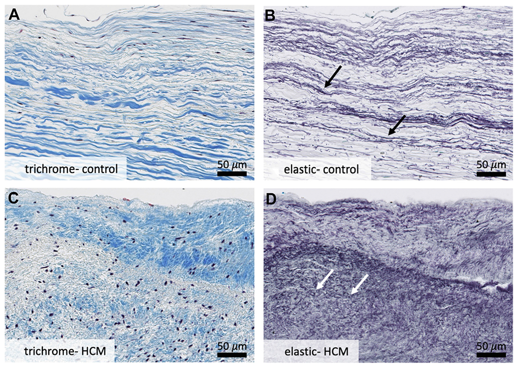
FIGURE 3. Specific Structural Changes.

Higher-powered analysis of leading edge of control mitral valves (A, B) and hypertrophic cardiomyopathy residual leaflets (C, D) reveals disruption of the collagenous fibrosa layer and increased proliferation and fragmentation of elastic fibers in hypertrophic cardiomyopathy valves (D). A layer of superimposed collagenous tissue is also noted on surface of hypertrophic cardiomyopathy valves (C) that is not present on control valves (A). White arrows point to short, truncated elastic fibers as compared to normal fibers indicated by black arrows above. Overall, these changes are consistent with chronic hemodynamic stress on the hypertrophic cardiomyopathy residual leaflet. Weigert Resorcin/Fuchsin, elastic fibers = black. Trichrome, collagen = blue. HCM = hypertrophic cardiomyopathy.
