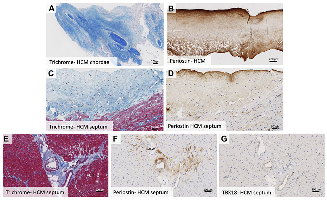
FIGURE 4. Notable Histologic Features.

In some hypertrophic cardiomyopathy subjects, superimposed tissue extended beyond the valve, encasing the chordae tendineae at their insertion sites (A). Hypertrophic cardiomyopathy leaflets featured patchy periostin staining of the atrial and ventricular valve surfaces and with areas of superimposed tissue staining strongly (B). Endocardial surface fibrosis with neovascularization, lymphocyte proliferation, and periostin staining is noted in septal myocardium samples (C, D). Septal myocardial samples in hypertrophic cardiomyopathy patients showed interstitial and replacement fibrosis, with abnormal intramyocardial vessels with perivascular fibrosis that stained positive for periostin (E, F). Many cells comprising abnormal arteries exhibited Tbx18 positivity (G). HCM = hypertrophic cardiomyopathy; Tbx18 = t-box transcription factor 18.
