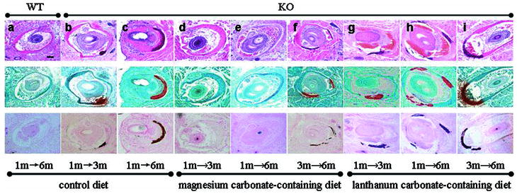
Figure 1. Histopathologic evaluation of mineralization of the vibrissae in Abcc6-/- mice (KO) or their corresponding wild-type (WT) counterparts kept on control, magnesium carbonate-containing, or lanthanum carbonate-containing diet.
The mice were placed on the experimental diet either at one month or three months of age, and biopsies from the muzzle skin containing the vibrissae were taken at three or six months of age. The tissue specimens were processed and stained with hematoxylin and eosin, Alizarin Red or von Kossa stains (top, middle, and bottom panels, respectively). All figures have the same magnification: bar, 100 μm.

