Figure 4.

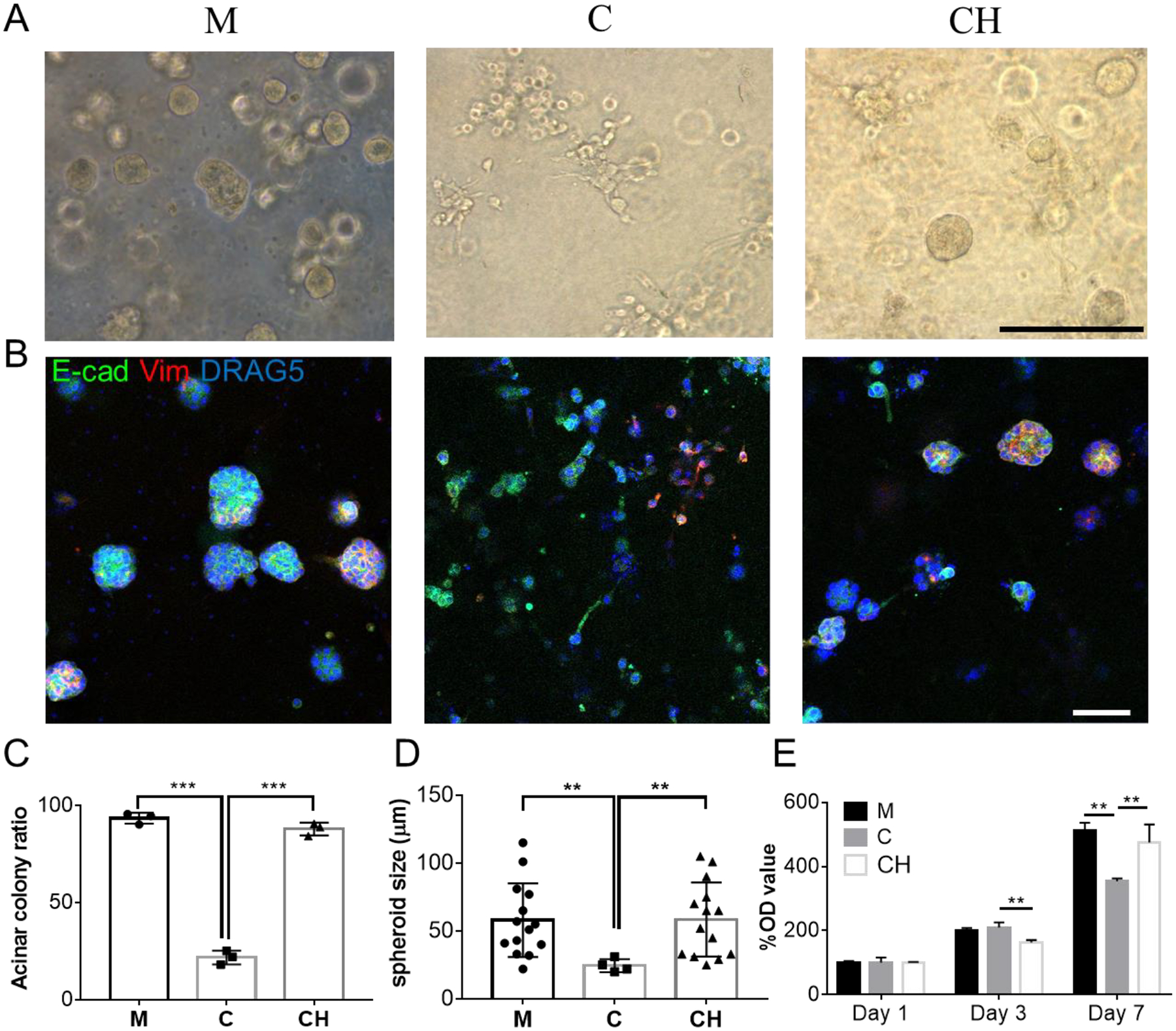
Behavior and morphology of 21PT human breast cancer cells in three models with different bioinks: Matrigel (M), collagen (C) and CH at day 7. A) 21PT cells formed mostly non-invasive acinar colonies in Matrigel and CH models but mostly non-acinar colonies in the collagen model. B) IF staining of E-cadherin (E-cad, green) and vimentin (Vim, red) proteins on 21PT cells in three bioprinted models. C) Quantification 21PT cells forming the acinar colony in three bioprinted models (n=3, ***p<0.001). D) Comparison of spheroid size formed in three bioinks (n=14, **p<0.01). E) Proliferation of 21PT cells in three bioprinted models determined by MTT study (n=4, **p<0.01; NS, not significant). Scale bar: black, 250 μm; white, 100 μm
